WikiJournal of Medicine/Medical gallery of David Richfield 2014
![]()
WikiJournal of Medicine
Open access • Publication charge free • Public peer review • Wikipedia-integrated
This article has been through public peer review.
First submitted:
Accepted:
Reviewer comments
PDF: Download
DOI: 10.15347/wjm/2014.009
QID: Q44276803
XML: Download
Share article
![]() Email
|
Email
| ![]() Facebook
|
Facebook
| ![]() Twitter
|
Twitter
| ![]() LinkedIn
|
LinkedIn
| ![]() Mendeley
|
Mendeley
| ![]() ResearchGate
ResearchGate
Suggested citation format:
David Richfield (27 July 2014). "Medical gallery of David Richfield 2014". WikiJournal of Medicine 1 (2). doi:10.15347/WJM/2014.009. Wikidata Q44276803. ISSN 2002-4436. https://upload.wikimedia.org/wikiversity/en/1/13/Medical_gallery_of_David_Richfield_2014.pdf.
License: ![]()
![]() This is an open access article distributed under the Creative Commons Attribution License, which permits unrestricted use, distribution, and reproduction, provided the original author and source are credited.
This is an open access article distributed under the Creative Commons Attribution License, which permits unrestricted use, distribution, and reproduction, provided the original author and source are credited.
Mikael Häggström ![]() (handling editor) contact
(handling editor) contact
Article information
Abstract
Gallery
-
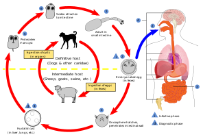 Life cycle of Echinococcus
 -
 A diagram of a myofibril
 -
 A diagram of the movement of the units of a myofibril
 -
 Anatomical planes, including median (red), parasagittal (yellow), frontal or coronal plane (blue) and transverse or axial plane (green).
 -
 Anatomical planes through a model of a human brain with eyes but excluding optic nerves, including median (red), parasagittal (yellow), frontal or coronal plane (green) and transverse or axial plane (blue)
 -
 Anatomical planes, including median (red), parasagittal (yellow), frontal or coronal plane (green) and transverse or axial plane (blue)


