Upper Limb Orthotics/Wrist Contracture Management
Click edit and copy the text from here to paste on your Case Study page
Case Study Description
editThe patient is a 12 year old male who had sustained a burn injury on the palmar aspect of his hand. The patient accidentally placed his hand on the barbeque plate during an outing with his family. The patient was treated at the hospital for 3rd degree burn. Skin grafting was performed to his thenar eminence, hypothenar and first web space of the burned hand. After the surgery, patient would be required to maintain his hand in neutral position to facilitate healing and to prevent contracture.
Evidence
editChildren are more susceptible to burn injuries especially on their hands. This may be associated to their natural curiosity, skin sensitivity and slow withdrawal reflexes (Yotsuyanagi, Yokoi & Omizo, 1994). Third-degree burns are very serious as it involves epidermis, dermis, subcutaneous tissue and even the muscles tissue (Howell, 1989). Therefore, third-degree palmar burned injuries in children are managed in a careful and systematic way to ensure optimal functionality of the region is achieved. Successful management of the burned hands does not result simply from closure of the wound (Moore, Dewey & Richard, 2009). It involves extensive care from multidisciplinary teams for a period of time to prevent deformity and restore function. Surgical intervention is usually performed to minimise further complications, restore function and increase range of motion (Sabapathy, Bajantri, Bharathi, 2010). Third-degree palmar burn require skin grafting to be performed as soon as possible, usually within 72 hours of injury (Sheridan, Baryza, Pessina, O'neill, Cipullo, Donelan, Ryan, Schulz, Schnitzer, & Tompkins, 1999). Otherwise, the burned palmar of the hands can lead to flexion contracture and loss of first web space. Flexion contracture can cause deformity that can affect the functionality of the hand. The loss of first web space can compromise the use of the thumb and cause deformity. Therefore, orthotic management is crucial part of the rehabilitation to facilitate healing and most importantly to prevent contracture to the carpometacarpal joint, metacarpal phalangeal joint and interphalangeal joint.
Pathophysiology of contracture
editThe hand is rated the third most frequent site for burned scar contracture (Moore, Dewey & Richard, 2009). Deformity due to contracture is prevalent with children who underwent poorly managed third-degree burned injuries (Sykes, 1991). Burn contracture progresses slowly throughout the healing process. The skin is slowly replaced by non-functional mass of tissues, known as scar tissues. Burn contracture is a late event of secondary healing when the mature scar thickens and tightens, causing contracture to the healing tissue. According to Yotsuyanagi, Yokoi & Omizo (1994), hypertrophic scar contracture is commonly seen in third-degree burn tissues and remains conspicuous after healing. Contracture also happens when there is a prolong immobilization of the joint in an inappropriate position (Dewey, Richard, Parry, 2011). Contracture produces deformities that result in permanent loss of functional loss of hand and also stigma disfigurement. (Bhattacharya, 2013; Farmer, Woollam, Patrick, Roberts, & Bromwich, 2005). Contracture on the palmar aspect of the hand causes stiffness to the wrist joint, ligaments, muscles and skin.
Position of contracture
editMaintaining the palmar burned hand is important because the position of the hand has been highly associated with flexion contracture. Children usually adopt themselves to a comfortable position where wrist joint is slightly flexed and radial deviated. As the healing takes place, the range of motion of wrist joints, metacarpophalangeal joints and interphalangeal joints will quickly reduce to a non-functional position (Procter, 2010). This is known as the position of deformity where clinical presentation will show radial deviation, flexion of the wrist joints, extension of the metacarpal joints, hyperextension of the forth and fifth metacarpophalengeal joints, and flexion of the proximal and distal interphalangeal joints (Bhattacharya, 2013; Gendron, Jain & Delisa, 1992; Howell, 1989). In addition, it will also show loss of transverse metacarpal arch, thenar eminence approaching hypothenar eminence and adducted thumb position (Sabapathy, Bajantri & Bharathi, 2010). This deformity is also known as the palmar cupping deformity where the grasping surface of the hand is lost (Procter, 2010).
Orthotic treatment options
editOrthotics is used throughout the burned rehabilitation to immobilize the joint and to facilitate healing. Burned injuries in children should be address immediately with orthosis and use through all phase of rehabilitation for better outcome. Orthotics is best to use from the start of the treatment to prevent the development of post-burn scar contracture as opposed to correcting the contracture once it has occurred. Studies have shown good outcomes when using orthotics to manage third-degree burn patients. Huang, Blackwell & Lewis (1978) stated in their studies that incidences of contractures and frequencies of secondary surgeries to release contracture have decrease remarkably for patients using orthotics for at least 6 months. Sheridan et al (1999) also concluded in their study that excellent functional results are achievable if the burned hand is managed systematically with orthotic.
Orthotic principles
editOrthotics uses biomechanics principles to maintain joint positions or to effectively apply forces to correct certain joints to ideal positions. Orthotic designs are based on three simple principles. The three principles are pressure applied to large areas principles, equilibrium principles and lever arm principles (Duncan, 1989). Using correct techniques is crucial in managing the contracture. Otherwise, the orthotics can cause more damage than good such as creating localized pressures or even contractures to other joints. If the orthotics is used to correct contracture, the force applied to the area of interest must not be too much or too little. If the force applied is too much, it can damage healing tissues. On the other hand, if the force is too little, it will not stimulate change in the tissues (4).
Orthotic and tissues biomechanics
editTissues react in different ways in order to adapt the mechanical force from the orthotics. The processes of the tissue change are tissue stress relaxation and tissue creep (Dewey, Richard & Parry, 2011). Tissue stress relaxation happens when the orthotic is fitted to burn patient with their tissues under tension. However, the tension on the tissues is only temporary because it eventually subsides to a more comfortable level due to elongation changes in cellular levels in response to the tension (Dewey, Richard & Parry, 2011; Procter, 2010). Similarly, tissue creep goes through the same cellular level changes as tissue stress relaxes. But, tissue creep happens when an orthotic applies a constant force to gradually elongate the tissue over a period of time (Farmer, Woollam, Patrick, Roberts & Bromwich, 2005). Farmer & Woolam stated that when connective tissues are held at a constant length for a period of time, the tissues would slowly relax. Therefore, orthotics used to hold any joint in a fixed position will eventually lose it stretching effect.
Orthotic positioning
editUnderstanding of the position of deformity is fundamentally important so that the orthotics can minimize or prevent the contracture from happening. Most of the literature has different suggestions for the anti-deformity position. However, the suggestion of the anti-deformity position is subjective, as the extent of the damage to the soft tissues have to be considered. It have been suggested that the ideal position for the palmar burned hand is 15 to 30 degree wrist extension, 60 to 90 degree metacarpophalangeal joint flexion and fully extended interphalangeal joint. The thumb should be positioned at full abduction and extension with the arches of the hand preserved (Gendron, Jain & Delisa, 1992; Howell, 1989; Moore, Dewey & Richard (2009). This position is also known as the open palm position where it provides maximum palmar stretch and is beneficial in preventing shortening of the collateral ligaments (Hsu, Michael & Fisk, 2008).
Comparison of orthotic treatment options
editThere are various types of orthotics that can be used in burn management such as static orthotics, dynamic orthotics and static progressive orthotics. Static orthotics is often used to maintain a fixed position after the burned hands have underwent surgeries or to maintain the hand at an anti-deformity position (Cowan & Stegink-Jansen, 2013). As the static orthotics maintains a fixed position, it needs to be periodically reviewed to ensure the tissue is not under stretched. Both static progressive orthotics and dynamic orthotics are often used to correct contracture of the burned hand by creating tension to the tissues so that the tissues can adapt to the tension, via stress relaxation and tissues creep. Static progressive orthotics requires tension to be manually adjusted each time the joint reaches the ideal position (Schultz-Johnson, 2002). Conversely, dynamic orthotics does not require manual adjustment as it has a dynamic constant force to the joints to correct the range of motion. However, dynamic orthotics is not recommended for children as the small parts used in the orthotics pose a hazard to them.
Functional Aims and Goals
editTraditionally, plaster of paris (POP) cast was used to manage burned hands. As burned hand requires continuous dressing and antibiotic cream treatment, POP cast has since lost its popularity to thermoplastic orthosis. However, the functional aims and goals for both POP cast and static orthosis in burn management remain the same. It is used to immobilize wrist joints, carpometacarpal joints and interphalangeal joints. Besides that, it also acts as a protection barrier to the burned and grafted area. When using static orthosis, wrist joint, carpometacarpal joints, and interphalangeal joints are immobilize and stretch progressive to prevent flexion contracture. This enables healing to take place without causing deformity to the bones, ligaments, tendons and surround tissues. Apart from that, the thumb and fingers are also abducted to stretch and preserve web spaces. Web spaces are crucial in allowing the hand to function in different plane. On the other hand, due to the damage on the epidermis layer, the burned area losses its protection layer from pathogens. Static orthosis minimise the risk of infection by acting as a protection layer between the burned area and the outer environment. It also helps to protect the newly grafted area from infection and external trauma.
Design & Positioning
editThe design of the orthosis (Figure 1) consist of the following features
- Prevention of flexion contracture of the wrist joint, metacarpophalangeal joints and interphalangeal joints
- Holding all digits fully abductor to preserve all web spaces
- Protection of the burned area
-
 Figure 1: Design of the orthosis

Trimlines should be extending from the distal digits to 2/3 of the forearm and cover half the circumference of the forearm. In this case, a low temperature plastic will be used to fabricate the device. Appropriate force is to apply to patient’s hand to hold the hand in desire position (Figure 2).
-
 Figure 2: Force diagram

Manufacturing process
editTools needed to manufacture the wrist hand orthosis:
- Plain cloth to draw pattern
- Towel
- Electric frypan
- Velcro loop and hook
- Heatgun
- Scissors
- Low temperature thermoplastic
Guidelines
1) Place the plain cloth underneath the hand of the patient (Palm face up and all digits fully abducted) 2) Trace along the hand and mark CMC joints and wrist joint 3) Add two centimeters along the trace line and four centimeters medial and lateral to the 1st and 5th digit of the template 4) Cut the pattern out and apply to the patient’s hand to check for fit. Adjust trimlines if too excessive and redraw if the trimlines are not sufficient (Figure 3).
-
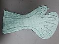 Figure 3: Trace pattern of the hand and trimlines

5) Next, cut thermoplastic according to the final template. 6) Position patient to an appropriate position. Palmar surface of the hand should face upward (Figure 4) with all digits fully abducted on the corner of the table (Figure 4 & 5). This position allows you to utilize the gravity force to position the thermoplastic on patient’s hand.
-
 Figure 4: Positioning of the hand
 -
 Figure 5: Positioning of the hand


7) Once patient is ready and all tools are set up, submerge the low temperature plastic into boiling water and leave to heat. 8) Then, remove the low temperature plastic and place on top of the towel to remove excess water. 9) Mould the low temperature plastic to patient’s forearm and hand. Make sure the plastic is push between all web spaces of the digits (Figure 6, 6a & 6b)
-
 Figure 6: Positioning of the hand (with thermoplastic)
 -
 Figure 6a: Positioning of the hand (with thermoplastic)
 -
 Figure 6b: Positioning of the hand (with thermoplastic)


10) Remove the plastic when it is set and neatly trim the edge of the plastic to the desire trimlines. It is easier to heat the plastic in the hot water before cutting it off.
11) Cut a long piece of plastic approximately 3cm width to reinforce the palmar surface of the orthosis ((Figure 6& 6a) 12) Apple the Velcro hook, loop and straps to hold the hand and to achieve desire force system as per design (Figure 7&7a)
-
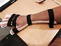 Figure 7: WHO orthosis with straps (Dorsal view)
 -
 Figure 7a: WHO orthosis with straps (Anterior view)


Critique of fit
edit-
Critique video
Outcome measures
editOutcome measures such as range of motion (ROM) testing, muscle grading and The Disability of the Arm, Shoulder and Hand Questionaire (DASH) were used to evaluate this patient.
The ROM of wrist joint, MCP, and IP were tested periodically to ensure adequate range of motion is still preserved. Patient is able to achieve satisfactory range of motion to perform daily activities. Patient losses some range of motion as a result of muscles stiffness. ROM revealed patient is able to achieve 65 degrees flexion of the wrist joint, 40 degrees flexion of thumb MCP joint, 70 degrees flexion of the thumb IP joint, 70 degrees flexion of the fingers MCP, 90 degrees flexion of the finger PIP joint and 60 degrees flexion of the finger DIP joints.. Muscles grading was performed to the hand and received a score of 4, where is could be moved against gravity and some resistance. DASH was used to evaluate if patient is able to perform certain activities. The DASH evaluates the upper limb disorder in terms of both function and symptoms using a score of 100, where higher numbers attribute to higher disability (Institute for Work & Health, 2013), Patient score 30 on this assessment, indicated patient is at the threshold for discharge from treatment and aware the limitation of their limb but did not consider them a problem. However, patient is advice to continue physiotherapy in order to achieve full range of motion.
Therefore, it is concluded that the use of the WHO orthosis is useful in preventing flexion contracture of the burned palmar hand. Patient is able to achieve satisfactory range of motion. Besides that, patient is able to perform his daily activity with minimal restriction.
References
editAarabi, S., Longaker, M. T., & Gurtner, G. C. (2007). Hypertrophic scar formation following burns and trauma: new approaches to treatment. Plos Medicine, 4(9), 1464-1470. doi: 10.1371/journal.pmed.0040234
Bhattacharya, S. (2013). Avoiding unfavorable results in postburn contracture hand. Indian Journal Of Plastic Surgery, 46(2), 434-444. doi: 10.4103/0970-0358.118625
Cowan, A. C., & Stegink-Jansen, C. W. (2013). Rehabilitation of hand burn injuries: Current updates. Injury, 44(3), 391--396. doi: 10.1016/j.injury.2013.01.015
Dewey, W. S., Richard, R. L. & Parry, I. S. (2011). Positioning, splinting, and contracture management. Physical Medicine And Rehabilitation Clinics Of North America, 22(2), 229-247. doi:10.1016/j.pmr.2011.02.001
Duncan, R. M. (1989). Basic principles of splinting the hand. Physical Therapy, 69(12), 1104-1116. Retrieve from: http://www.ncbi.nlm.nih.gov/pubmed/2587634
Farmer, S.E., Woollam, P.J., Patrick, J.H, Roberts, A.P, & Bromwich, W. (2005). Dynamic orthoses in the management of joint contracture. Journal Of Bone & Joint Surgery, British Volume, 87-B(3), 291-295. doi: 10.1302/0301-620X.87B3.
Gendron, B. C., Jain, S. S., & Delisa, J. A. (1992). Burn Care and Rehabilitation. Indian Journal Of Physical Medicine & Rehabilitation, 5(1), 1-9. Retrieve from: http://www.ijpmr.com/ijpmr92/199201 Howell, J.W. (1989). Management of the acutely burned hand for the nonspecialized clinician. Physical Therapy, 69(12), 1077–1090. Retrieve from: http://ptjournal.apta.org/content/69/12/1077 Huang, T. T., Blackwell, & S.J., Lewis, S.R. (1978). Ten years of experience in managing patients with burn contractures of axilla, elbow, wrist, and knee joints. Plastic And Reconstructive Surgery, 61(1), 70-76.
Hsu, J. D., Michael, J. W. & Fisk, J. R. (2008). AAOS atlas of orthoses and assistive devices. Philadelphia: Mosby/Elsevier.
Institute for Work & Health. (2013). The DASH outcome measure - Disabilities of the Arm, Shoulder and Hand. Retrieved from http://www.dash.iwh.on.ca
Moore, M. L., Dewey, W. S., & Richard, R. L. (2009). Rehabilitation of the burned hand. Hand Clinics, 25(4), 529-541. doi: 10.1016/j.hcl.2009.06.005
Procter, F. (2010). Rehabilitation of the burn patient. Indian Journal Of Plastic Surgery, 43(0), 101-113. doi: 10.4103/0970-0358.70730
Sabapathy, S. R., Bajantri, B., & Bharathi, R. R. (2010). Management of post burn hand deformities. Indian Journal Of Plastic Surgery: Official Publication Of The Association Of Plastic Surgeons Of India, 43 (0), S72-S79. doi: 10.4103/0970-0358.70727
Schultz-Johnson, K. (2002). Static progressive splinting. Journal Of Hand Therapy, 15(2), 163-178. Retrieve from: http://0-search.proquest.com.alpha2.latrobe.edu.au/docview/222231341?accountid=12001
Sheridan, R. L., Baryza, M. J., Pessina, M. A., O'neill, K. M., Cipullo, H. M., Donelan, M. B., Ryan, C. M., Schulz, J. T., Schnitzer, J. J., & Tompkins, R. G. (1999). Acute hand burns in children: management and long-term outcome based on a 10-year experience with 698 injured hands. Annals Of Surgery, 229(4), 558-564. Retrieve from: http://www.ncbi.nlm.nih.gov/pmc/articles/PMC1191743/?tool=pmcentrez&report=abstract
Sykes, P. (1991). Severe burns of the hand: a practical guide to their management. The Journal Of Hand Surgery, 16(1), 6-12. Retrieve from: http://www.ncbi.nlm.nih.gov/pubmed/2007816
Yotsuyanagi, T., Yokoi, K., & Omizo, M. (1994). A simple and compressive splint for palmar skin grafting in young children with burns. Burns, 20(1), 55-57. Retrieve from: http://dx.doi.org/10.1016/0305-4179(94)90107-4
Search Strategies
editSearch strategies was used in search databases to review the managemnt of the burned contracture. The databases used in searching for relevant articles are CINAHL, Cochrane and Medline. Soem articles was obtained through a general search using Latrobe Libguides and Google Scholar. In particular, all searches use key words such as palmar burned contracture, splinting, orthosis, third degree burn, full thickness burn, and orthotic management.