Upper Limb Orthotics/Smith's Fracture
Describe your case study
editThe patient is a 24 year old male who suffered an injury playing AFL. He is a professional AFL Footballer fighting for his position in the squad, the injury occurred in a match when he was unexpectedly pushed in the back, he fell on top of his right hand in a flexed position which resulted in a distal radius fracture with volar displacement of the distal fragment (Smith’s Fracture). There is a significant ulnar protrusion and the normal anterior curve of the distal radius head is extremely exaggerated. The patient has undergone current reduction and is in a cast however, the fracture is unstable and has become unaligned. Using the modified Thomas classification system, the client has a type 2 Smith’s fracture. The patient would like to get back to training and playing football as quick as possible to be able to maintain his position in the squad.
Written information
editEvidence
editAnatomy & Pathology: A Smith fracture is a distal radius fracture, commonly referred to as a reverse Colles’ fracture, which has volar displacement of the distal fragment. Distal radius fracture are the most common upper limb fracture and constitute 8% to 15% of all bone injuries (Hanker,2001).
A smith’s fracture occurs at the distal radius, it can have an impact on the wrist joint stability, involving the proximal row of carpals and articular surfaces of the radius, and surrounding tendons such as extensor pollicis longus. The distal radius has “two articular surfaces, the radiocarpal joint with the scaphoid and lunate facets, and the sigmoid notch that articulates with the distal ulna.” (Capo, Swan & tan, 2006). The smith fracture can include these articular surfaces and can lead to further complications. Both these articular surfaces need to be realigned to allow for optimal function (Capo,Swan & Tan, 2006). Involvement of these articular surfaces in the fracture can lead to instability of the fracture and a likely chance of displacement to reoccur after reduction.
The fracture can have an impact of our mobility of our fingers especially our thumb. In the literature it has been well documented that “tendon damage may occur as a complication of distal radius fractures”(Murakami & Todani, 1981). The position of the fracture being at the distal end of the radius allows it to become a problem when reduction occurs with a chance of “entrapment of the extensor pollicis longus” (Itoh et al., 1987). This complication can cause stiffness and pain (Kelly & Kumar, 2002) for the client and can lead to further surgery.
This specific type of distal radius fracture is quite rare compared to the Colles’ fracture as it is uncommon for an individual to fall on their flexed hand. The fracture can occur in multiple different ways; it is usually a result from “a fall on the dorsum of the hand” (Jacobs & Austin, 2013) however another concept of how the fracture can occur, as stated by Flandreau, Sweeney and O'Sullivan (1962), is the individual falling backwards on their outstretched supinated hand causing volar displacement of the distal fragment as the proximal fragment is forcibly pronated while the hand is locked in a supinated position on the ground combined with compressive forces. Picture here in fig 14 & 15
Smith fractures can be categorized using a modified Thomas classification system (Woodyard, 1969):
Type 1) extra-articular.
Type 2) Fracture line crosses into the dorsal articular surface.
Type 3) Fracture line enters the carpal joint.
To see examples here in fig 2.17
Orthotic treatment options
edit(this may include post-surgery)
The best treatment of a fracture is one that “should restore and maintain anatomy, allow early mobilisation and have minimal risk of complications” (Fang, Kwan & Leung, 2013). The type of orthotic treatment of a Smith’s fracture is dependent on the severity, classification type, of the fracture and whether the fracture is going to be operated on or not.
If surgery is not going to be under taken for the client, a below elbow cast will be placed on the client after closed reduction. This immobilisation technique will “hold the wrist in slight extension, the forearm in full supination, and the elbow at 90 degrees of flexion” (Fernandez & Jupiter, 2002) with a below elbow sugar tong cast. If the fracture is extra-articular the hand will be placed in 20 to 30 degree extension as mentioned above, if it is an intra-articular fracture and non-operative treatment is attempted it is recommended to be placed in slight flexion to “maintain congruity between the proximal row of the carpus and the intact part of the lower end of the radius.” (Thomas, 1957). Picture of this concept here in fig 16 and 17. The cast should remain on for 6 weeks (Fernandez & Jupiter, 2002). With a removable splint used for the following weeks.
Dependent on the surgery undertaken, different post-surgery orthotic interventions can be used. The different types of surgery involve non-bridging external fixation, bridging external fixation, Kirschner-wires or internal fixation with volar placed buttress plate. According to Fernandez & Jupiter percutaneous k-wires used for an intra-articular compression fracture should use a thumb spica splint for post-surgical recovery. Whereas Michlovitz, Lestayo, Alzner and Watson (2001) states that a splint that allows all metacarpal and intercarpal range of motion of the digits is recommended for a quicker recovery, such as a keyhole wrist hand orthoses.
Comparison of orthotic treatment options
edit(or orthotic treatment vs surgical intervention)
As mentioned before there are multiple surgical interventions. These surgical interventions should be used only when the joint is unstable and a high risk of displacement. According to Nazar,Mansingh, Bassi and Waseem (2008) there is not a set management technique used for distal radius fractures like a smiths fracture, a magnitude of different treatment plans are utilised and are chosen upon dependent on the individual’s needs, goals and severity of injury. However, Jacobs and Austin (2013) believe there is an increase of incidence of volar plated management of distal radius fractures due to improvement of the plating designs.
In a randomly controlled trial of distal radius fractures in 56 individuals comparing volar plates and k-wires treatment success conducted by Mcfadyen et al. (2011) the volar plate had a quicker return to functionality and posted better scores then k-wires in DASH tests. The volar plates created no complications however, 28% of the clients treated with K-wires contracted a complication of sort, such as pin site infection or carpal tunnel syndrome occurring. In relation to the previous literature it seems the volar plate internal fixation leads to a quicker return from injury, a low risk of complications and re-displacement and further surgical requirements in the future in relation to the fracture and k-wire treatment. In agreement, Shyamalan, Theokli, Pearse and Tennent (2009) mention “volar locked plat fixation provides a more stable fixation and allows an earlier range of wrist motion then percutaneous fixation”.
External fixation is a well-known treatment for distal radius fractures like the smith’s fracture. Although, in a study comparing external fixation and volar plating conducted by Koshkin, Sergeev, Matveev and Grishanin (2008) the results show volar plating being a greater treatment option than a bridging external fixation such as the motube triax device (stryker) that was used in the study. The plating treatment resulted in 15 out of 22 patients ending in a good result compared to 11 out of 32 in the external fixation group, results recorded using E. R. Mattis' score system. In agreement, Murray and Trigg (2002) state sustained longitudinal traction created by external fixation can lead to increased rates of complications.
Summary
editAs the client has had “redisplacement in [a] plaster support, [that] defines the fracture as being unstable, and that an alternative approach to maintain the reduction should be considered” (Fernendaz & Jupiter, 2002) it is therefore in the clients best interests to undertake surgery for his fracture. With limited literature on the post operational orthotic devices recommended for Smith fracture treatment, it is recommended for the client to undertake the surgery that allows for a quick recovery and allow for early mobilisation and has a low risk of complications. In review of the literature the client shall undertake a volar plate internal fixation, resulting in an above elbow cast with the hand in neutral position for two weeks and then replaced with a removable keyhole wrist hand orthosis with the hand only being used in daily living activities for the first 6 to 8 weeks (Fernandez & Jupiter, 2002) this treatment is allowing for early movement, non-contact training to be undertaken and extremely light hand skills such as handballing and short kicking. In case of swelling and sensitivity over the surgical site, volar distal radius surface, pressur is released from that area. After 12 weeks the client should be back playing competitively.
Reference
editCapo, J. T., Swan Jr, K. G., & Tan, V. (2006). External fixation techniques for distal radius fractures. Clinical orthopaedics and related research, 445, 30-41.
Cooper, C. (2013). Fundamentals of Hand Therapy: Clinical Reasoning and Treatment Guidelines for Common Diagnoses of the Upper Extremity: Elsevier - Health Sciences Division.
Fang. C., Kwan. K., & Leung. F. (2013) Focus on: Distal Radius Fracture: Current concepts and management. Bone & Joint. Retrieved from http://www.boneandjoint.org.uk/sites/default/files/Distal%20radius%20fracture.pdf
Fernandez, D. L., & Jupiter, J. B. (2002). Fractures of the Distal Radius: A Practical Approach to Management: Springer New York.
Flandreau, R. H., Sweeney, R. M., & O'Sullivan, W. D. (1962). Clinical experiences with a series of Smith's fractures. Archives of Surgery, 84(3), 288.
Hanker, G. J. (2001). Radius fractures in the athlete. Clinics in sports medicine, 20(1), 189-201.
Itoh, Y., Horiuchi, Y., Takahashi, M., Uchinishi, K., & Yabe, Y. (1987). Extensor tendon involvement in Smith's and Galeazzi's fractures. The Journal of hand surgery, 12(4), 535-540.
Jacobs, M. L. A., & Austin, N. M. (2013). Orthotic Intervention for the Hand and Upper Extremity: Splinting Principles and Process: Wolters Kluwer Health.
Jupiter, J. B. (1991). Current concepts review: fractures of the distal end of the radius. J Bone Joint Surg Am, 73(3), 461-469.
Koshkin, A. B., Sergeev, S. V., Matveev, V. S., & Grishanin, O. B. (2008). Distal forearm fractures: the analytical approach for treatment. Ortop Traumatol Rehabil, 10(4), 324-30.
Kumar, A., & Kelly, C. P. (2003). Extensor pollicis longus entrapment after Smith’s fracture. Injury, 34(1), 75-78. Liporace, F. A., Adams, M. R., Capo, J. T., & Koval, K. J. (2009). Distal radius fractures. Journal of orthopaedic trauma, 23(10), 739-748.
McFadyen, I., Field, J., McCann, P., Ward, J., Nicol, S., & Curwen, C. (2011). Should unstable extra-articular distal radial fractures be treated with fixed-angle volar-locked plates or percutaneous Kirschner wires? A prospective randomised controlled trial. Injury, 42(2), 162-166.
Michlovitz, S. L., LaStayo, P. C., Alzner, S., & Watson, E. (2001). Distal radius fractures: therapy practice patterns. Journal of Hand Therapy, 14(4), 249-257.
Mostofi, S. B. (2013). Fracture Classifications in Clinical Practice 2nd Edition: Springer.
Murakami, Y., & Todani, K. (1981). Traumatic entrapment of the extensor pollicis longus tendon in Smith's fracture of the radius—Case report. The Journal of hand surgery, 6(3), 238-240.
Murray, P. M., & Trigg, S. D. (2002). Treatment of distal radius fractures with external fixation: technical considerations for rehabilitation. Techniques in hand & upper extremity surgery, 6(4), 213-218.
Nazar, M. A., Mansingh, R., Bassi, R. S., & Waseem, M. (2008). Is there a Consensus in the Management of Distal Radial Fractures?. The open orthopaedics journal, 3, 96-99.
Orbay, J. L., & Fernandez, D. L. (2002). Volar fixation for dorsally displaced fractures of the distal radius: a preliminary report. The Journal of hand surgery, 27(2), 205-215.
Shyamalan, G., Theokli, C., Pearse, Y., & Tennent, D. (2009). Volar locking plates versus Kirschner wires for distal radial fractures—a cost analysis study.Injury, 40(12), 1279-1281.
Thomas, F. B. (1957). Reduction of Smith's fracture. Journal of Bone & Joint Surgery, British Volume, 39(3), 463-470.
Woodyard, J. E. (1969). A review of Smith’s fractures. children, 3, 3.
Functional Aims and Goals
editThe Low Temperature Thermoplastic (LTT) cast has multiple functional aims and goals.
The design is too add support for the volar plate that has been surgical inserted over the fracture site to help with realignment and quick recovery. To help with this support the device’s trimlines are over the carpal joints and finish proximal to the distal palmar crease and distally from the elbow joint.
The device will immobilise the carpal joints and will hold the hand in slight extension, preferably between 20 – 30 degree, and with slight ulnar deviation. This will help maintain the alignment the volar plate is creating and keep promoting the healing process.
It will allow for movement of the phalanges and thumb at the metacarpophalangeal joints and carpometacarpo joint, so the client is still able to use that hand in light daily activities. The device will be removable.
The device is to be comfortable to wear and not to produce any rubbing or pain for the client, it is to conform to the clients hand and the trimlines are to be smooth. A pressure release over the surgical incision site, distal radius, will be ensured to reduce any pain and help the healing process of the wound.
The goals of the device to allow for a quick healing process and allow for early movement of the hand so the client can do very light exercises with the football and maintain his ball skills.
The POP cast designed share the same functional aims as mentioned above.
It is to allow for movement of the phalanges and thumb at the metacarpal joints. It will immobilise the carpal joint(wrist joint) in slight extension and ulnar deviation. It is to be comfortable to wear as well as ensure no rubbing over the sensitive skin of the surgical incision sit
Design
editThe chosen design for the Low temperature Thermoplastic cast was a Colles’ fracture brace.
This cast is to place the hand in 20-30 degrees of slight extension and slight ulnar deviation (10 degrees). This position will help with the alignment the volar plate has created and increase the healing process.
The cast is to allow movement at the metacarpophalangeal and carpometacarpal joint. Refer to Cast Design Below.
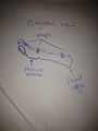
A 3 point force system is applied through this cast:
- Refer to 3 point force system figure below, F2 is a wrist extension force (applied at the dorsum of the wrist joint), F1 and F3 are forces opposing wrist extension (F1 applied through palmar surface, F3 through volar surface of the proximal end of the device)
The trimlines of the device should be contoured in a way that maximises comfort, stability and the healing process:
- End at distal palmar crease, edge is rolled back to minimise rubbing, and a few centre metres from the proximal end of the forearm to avoid pinching when flexion at the elbow joint occurs, this edge is flared. The cast should be 2/3 length of the forearm. The medial and lateral walls shall come up over halfway of the sides of the forearm. A hole shall be cut out to allow for thumb to fit through comfortably.
All straps wrap over the dorsal surface of the arm. They are located at the proximal end of the device, over the wrist joint and at the metacarpophalangeal joints. As shown in cast design figure 1 below. Straps will attach to sticky back Velcro loops.
-
 Cast Design
 -
 3 point force system


Manufacturing process
editPreparation phase
edit1) To create the pattern the use of a kitchen ‘Chux’ is ideal, have the patient seated and place their forearm and hand resting on the chux as shown in Fig 1, ensure fingers are close together. Draw an outline of the client hand.
2) Once outline is drawn mark the distal palmar crease, the first and second proximal interphalangeal joint & the matacarpophalangeal joint of the thumb. Now draw a horizontal line from the metacarpophalangeal joint of the thumb and a verticle line through the middle of the index finger which is identified form earlier markings. At the intersection draw an oval that is 1 cm wide and 2 cm long. Refer to Fig 2 & 3.
3) With a texta, now draw a pattern that is an inch wider medially and laterally then the forearm and cuts across at the distal palmar crease. It should cut through the thumbs metacarpophalangeal joint. Refer to Fig 3
4) Now cut this pattern out as well as the hole and fit it to the patient to see whether it is correct dimensions. Refer to Fig 4.
5) Once happy with pattern, trace it onto the Low Temperature Thermoplastic (LTT) with a pen. Place pattern into hot waterbath to soften it and allow the pattern to be easily and smoothly cut out. Refer to Fig 5.
6) Now to create a pressure release point in the cast, cut out a small rectangle slightly longer then the length of the cut/scar of the patient form the surgery, now stick this foam over the wound (do not have direct contact to wound but have a type of covering to avoid this) attaching it with tape Fig 4, Now cover the arm in a stocking tying the distal end, having a hole for the thumb and hinging the proximal end to the clients shoulder or clothes to make the stocking taught. Refer to Fig 6.
-
 Fig 1
Fig 1 -
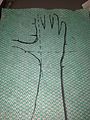 Fig 2
Fig 2 -
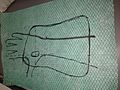 Fig 3
Fig 3 -
 Fig 4
Fig 4 -
 Fig 5
Fig 5 -
 Fig 6
Fig 6


Moulding process
edit7) While pattern is soft and in waterbath, place patient in correct position, wrist in 20-30 degree extension and 10 degree ulnar deviation with fingers relaxed. A goniometer can be used to help with angles. Ask patient to maintain correct position.
8) When pattern is ready, roll distal trimline and thumb hole before moulding cast.
9) Re-soften pattern, then while being careful the cast isn’t too hot place it on the clients forearm, putting the thumb through the hole first. While client maintains correct arm position, ensure all trimlines are correct. Now hold the clients hand in a ‘bikey’ hand grip to help it conform to the clients palm.
10) As the mould is nearly set, draw the final trimlines on the device and remove it from client. Cut the mould to desired size and flare proximal edge.
An example of moulding the cast can be found here: fracture LTT moulding.
Straps and Finishing Orthosis
edit1) Place device on the client and mark where Velcro loop pieces for straps and hook attachments. Cut out these pieces and get a heat gun.
2) Warm the sections of the orthosis where the Velcro hooks and loops are to be placed with the heatgun. Warming both the orthosis and back/sticky side of the velvro hook/loops and press these heated sides together. Positions shown in strap placement picture below.
3) Clean device and place on patient to check fit and to make sure there is no rubbing or rough edges on device. Adjust until you are pleased with the device fit.
-
 strap placement (sagittal view)

Critique of fit
editThe client is a 24 year old male who injured his right forearm playing AFL. On initial observation the patient presented with a significant ulnar protrusion and the normal anterior curve of the distal radius head was extremely exaggerated, a smith’s fracture was obvious. The client wishes to get back to training and playing as soon as possible.
Observation of the X-ray confirmed it was a type 2 smith’s fracture (Thomas classification system), a non-surgical approach was initially undertaken with casting. However, the bone became unaligned and so surgery was recommended. A volar plate was inserted to help with alignment and also as quick recovery is associated.
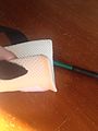
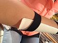
For rehabilitation a low temperature thermoplastic orthotic device was designed to allow for movement of the phalanges at the metacarpal joints,to keep the fracture aligned, to immobilise the wrist joint and not to rub on the wound site. The device was to hold the wrist in 20-30 degrees extension and 10 degree ulnar deviation. As shown in picture 1 and 2 below this was achieved. The device allows for movement at the metacarpal joints as what was prescribed, however as shown in picture 2 the thumb hole is quite large and I have stretched the material on the palmar surface. Taking more care in the thumb hole size in the designing phase and in the moulding process would stop this problem form occurring.
-
 Picture 1: 20-30 degrees of extension
 -
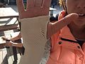 Picture 2: 10 degrees ulnar deviation


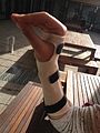
The pressure release over the wound is fine and allows for no rubbing to occur on the sensitive site as shown in picture 3. However, excessive flaring and lack of smoothing has caused the proximal trimlines of the device to be easily caught on sleeves or materials pictures 4 and 5. By cutting the corners to a more rounded edge, cutting the trimlines whilst the LTT is still warm will minimise the sharp or rough edges and finishing it all off with sandpaper will smooth the edges properly.
-
 Picture 3: release over surgical wound
 -
 Picture 4: excessive flaring
 -
 Picture 5: sharp corner


Using a texta in drawing the trimlines has caused it to smudge over the device and leave some unattractive marks, picture 6. The use of pencil would have been ideal when drawing trimlines, to avoid this ensure you are completely organised with everything you need before moulding the LTT to the client.
-
 Pictur 6: smudge marks from texta and rough trimlines

The distal trimlines are fine on the device, allowing for early movement at the metacarpal joints as shown in picture 7.
-
 Picture 7: allowing movement of the phalanges

Using the sport section in the DASH survey helps understand how the healing process is proceeding and functionality in the hand is going, which can be accessed here. However, the best outcome measure will be the patients strength and flexibility after 6 weeks and returning to training fully.
The client states the device is comfortable to wear and there is no rubbing at all from straps or over the sensitive wound site. The trimlines do not obstruct movement and he is able to perform majority of tasks with the device on and allows him to participate in very light training drills.
Outcome measures
editThe chosen outcome measure to be used was the Disabilities of the arm, shoulder and hand (DASH) questionnaire and Range of motion testing of the wrist joint. This was used as the DASH survey has a sport section which relates to my client perfectly. The range of motion tests were done as the clients joints are expected to be quite stiff when coming out of the cast and is required heavily in AFL.
The client was to answer the sport section before orthotic intervention and 6 weeks into orthotic intervention. The first time doing the survey was after surgery was completed. The questions asked and answers to these questions are in the table below.
| Question | 0 weeks | 6 weeks |
|---|---|---|
| using your usual technique for playing your sport? | 5 | 2 |
| playing your sport because of arm, shoulder or hand pain? | 5 | 2 |
| playing your sport as well as you would like? | 5 | 3 |
| spending your usual amount of time practising or playing your sport? | 5 | 1 |
(Questions extracted from the Disabilities of the Arm, Shoulder and Hand (DASH) survey)
As expected there was an increase in DASH results as his arm was broken and thus was unable to participate at all when initial measurements were undertaken. Although, after 6 weeks he is able to practice and train.
His right wrist is likely to be quite inflexible and lack a bit of strength as compared to his opposing wrist. The range of motion of his wrist was undertaken as well; checking flexion, extension, ulnar and radial deviation. Although was not done in initial consultation due to pain results were taken after 6 weeks.
| Type of Movement | Normal ROM | actual ROM |
|---|---|---|
| Flexion | 60 - 80 degrees | 53 degrees |
| Extension | 60 - 75 degrees | 67 degrees |
| Radial Deviation | 20 - 25 degrees | 16 degrees |
| Ulnar Deviation | 30 - 39 degrees | 27 degrees |
As expected the range of movement in the wrist was less than average due to stiffness. However, these results aren’t too extreme and should return to normal quite quickly with some physiotherapy sessions.
Referral Letter
editReferral was made to a sports physiotherapist to help with ROM and regaining strength of the wrist. It was addressed to: Mr. D. Farral - Sport Physiotherapist, Olympic Park Sports Medicine Centre (OPSMC), Olympic Boulevard AAMI PARK MELBOURNE, VIC 3004
Dear Mr. Farral,
Thank you for accepting my client referral,
The client is a professional AFL player; he is a 24 year old male who has suffered an injury in his dominant hand, right, whilst playing in a match. He suffered a distal radius fracture, Smith’s fracture, from falling on top of his flexed hand. The fracture was classified as a Type 2 smiths fracture according to Thomas classification system. Initial treatment was a non-surgical cast, however the fracture became unaligned and thus surgery was opted, volar plate internal fixation, with orthotic intervention for the healing process and rehabilitation.
The patient had an above elbow cast for two weeks post-surgery and then acquired a custom fitted removable low temperature thermoplastic (LTT) wrist hand orthosis to be worn as much as possible for 6-8 weeks.
The custom fitted LTT wrist hand orthosis allowed movement of the thumb and phalanges at the metacarpophalangeal joint. It immobilised the wrist joint to maintain the volar plate alignment and had a pressure release over the surgery wound to avoid any rubbing and complications with that healing process.
I am referring my client to you as he wishes to get back to AFL as quick as possible however, he has some slight stiffness and muscle weakness in the wrist joint and I was hoping you could provide your insight to help with the rehabilitation process for regaining that flexibility and strength as soon as possible.
I look forward to hearing your rehabilitation plans.
Your Sincerely,
Ned Verwey