Upper Limb Orthotics/Kienbocks Disease
-
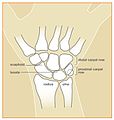 Fig 1. Bone Structure
 -
 Fig 2. Kienbocks x-ray


Describe your case study
edit- The patient is a 23 year old female who suffers from chronic wrist pain. She works as a deckhand on a cruise ship. She is constantly using her hands to lift anchors and throw ropes. Given that the patient has Kienbock’s disease and negative ulnar variance she has been referred for surgical intervention.
- The most comfortable resting position for the client is in 30 degrees of wrist extension, the digits are flexed to 50 degrees and the thumb is abducted to 40 degrees. On passive manipulation the wrist can be extended to 60 degrees and flexed to 40 degrees, the digits extended to 10 degrees and flexed to 55 degrees, and the thumb abducted to 55 degrees and the flexed to 85 degrees.
- The patient is currently undergoing treatment from an OT for referred forearm pain. The GP has requested an orthoses throughout her wait for surgery in 5/52.
- This device needs to prevent movement from the resting position. Requests to look simple and be comfortable for the patient to wear will be fulfilled.
Written information
editIntroduction
editKienbock’s Disease is defined as avascular necrosis of the lunate (Cross & Matullo, 2014, p. 141). The direct cause of Kienbock’s disease is largely unknown (Almquist, 1986, Nathan & Meadows, 1987) however, studies have confirmed an association between negative ulnar variance and the development of Kienbock’s Disease (Bonzar et al., 1998, Mirabello, Rosenthal, & Smith, 1987). Subjective information shows that ROM tests and palpation cause the patient a large amount of pain. It was identified that the most comfortable position for the client to be in is a supported, resting position. This is supported by Rizzo (2008, p. 491) by stating that avascular necrosis of the carpus is an important cause of wrist pain, limited motion, and weakness.
Evidence
editThe distal end of the radius and the articular disc of the distal radio-ulnar joint articulate with the proximal row of carpal bones (Moore, Dalley, & Agur, 2010, p. 809, para. 2). Therefore, on the proximal side of the lunate is the forearm and on the distal side is the second row of carpal bones. Given that these articular surface, the forearm and the wrist, are in connection with the lunate bone, any movements of the forearm may cause pain.
This pathology is common amongst manual workers or people lifting heavy loads repetitively. Repetitive trauma to the carpal bones, seen in manual workers, can lead to degenerative change in the lunate bone and can subsequently cause loss of blood supply to the bone (Laframboise, Gringmuth & Greenwood, 2012, p. 278).
A predictable pattern of arthritic degeneration and coinciding instability in the wrist can be evident as the disease progresses and the lunate collapses (UW Medicine The Hand Centre, 2009, para. 1). Therefore, given that there is no specific orthosis made for Kienbock’s disease, it is appropriate to design a resting orthosis, similar shape to that of chronic arthritis, to prevent any movement and pain.
The symptoms of Kienbock’s disease include; generalised or focal wrist pain, increased wrist stiffness and pain on flexion or extension, decreased grip strength, and limited range of motion (Alexander & Lichtman, 1986, Beckenbaugh, 1980, Laframboise, Gringmuth, & Greenwood, 2012). Laframboise et al., (2012) suggests that Kienbock’s disease presents with symptoms alike a sprain of the scapho-lunate ligament or a lunate fracture or dislocation.
Orthotic treatment options
editFrom the evidence it is clear that an orthosis for this patients injury needs to maintain an immobile wrist in a comfortable extension position. Any movement causes pain, subsequently it is appropriate to prescribe a rigid orthosis. Therefore, the Ottobock Manu Immobil Long (Ottobock, 2014, para. 1) is a prefabricated orthosis that would sufficiently immobilize the wrist before and after surgery. Gillen et al., (2008, p. 19) explained that pathologies including cumulative trauma disorders, arthritis, fractures, neurologic based weakness secondary to stroke, sprain, strain and tendonitis used wrist extension splints.
A similar custom device needs to be made of a rigid material and immobilize the carpal joints, the carpometacarpal joints, the metacarpophalangeal joints and the interphalangeal joints on the volar surface. This custom device can be fabricated from a HDPE thermoplastic including Orfit Eco or Technofit (Orfit, 2011, para. 11 & 14).
Comparison of orthotic treatment options
editIn line with the literature this patient had a period of resting and conservative treatment with an occupational therapist. An aged study by Agerholm and Goodfellow (1963, p. 111) found that a period of conservative treatment was incurred by most patients. In this study conservative treatment involved immobilization with plaster prior to operation, then if the conservative treatment failed it was usually an indication for operation. More recently, however, operation has been advised as soon as the diagnosis has been made (Agerholm & Goodfellow, 1963, p. 111).
Surgical correction presents several options. They include vascularised bone grafts (Rizzo, 2008, Matsuhashi et al., 2011), replacement of the bone with a prosthetic implant (Kato et al., 1986, p. 645) and radial shortening (Altay et al., 2008, p. 747).
A study by Dornan (1949, p. 518) assessed conservative treatment and surgical treatment. This study showed that conservative treatment of immobilization was provided to twenty-seven patients and surgical treatment was provided to sixteen patients. Of the conservative treatment patients, ten ended up have surgical treatment due to failure of conservative treatment. The results from this study found little difference between the results of conservative treatment and surgical treatment. This literature supports the idea that conservative treatment should be tried first, then surgical treatment can be assessed if conservative treatment fails.
Search Strategy
editThe databases used for the research in this paper were Cinahl and Google Scholar. Google was used as a general search engine for information. The keywords used to complete this search included; Kienbock, Kienbock AND treatment, wrist position AND splint, avascular necrosis, cause of Kienbock’s, Ottobock, Orfit, and negative ulnar variance. Ideas were extrapolated from indirectly related papers about the type of splint to use given that there is no definitive orthosis for this pathology. On evaluation, the sources were aged, but still valid references. Very limited amounts of new material is available on this pathology.
Conclusion
editIn conclusion, the patient, with Kienbock’s Disease and negative ulnar variance, is referred from her doctor to receive an orthosis to immobilize her wrist. After conservative treatment has failed the patient is waiting for surgery and requires a rigid, full-resting orthosis. This orthosis will be made out of a low temperature thermoplastic and will immobilize all joints of the wrist, hand and fingers. More recent research is needed for this pathology.
References
editAgerholm, J. C., & Goodfellow, J. W. (1963). Avascular necrosis of the lunate bone treated by excision and prosthetic replacement. Journal of Bone & Joint Surgery, British Volume, 45(1), 110-116.
Alexander, A. H., & Lichtman, D. M. (1986). Kienbock's disease. The Orthopedic clinics of North America, 17(3), 461-472.
Almquist, E. (1986). Kienbock's disease. Clinical orthopaedics and related research, 202, 68-78.
Altay, T., Kaya, A., Karapinar, L., Ozturk, H., & Kayali, C. (2008). Is radial shortening useful for Litchman stage 3B Kienbock’s disease?. International orthopaedics, 32(6), 747-752.
Bonzar, M., Firrell, J. C., Hainer, M., MAH, E. T., & McCabe, S. J. (1998). Kienböck Disease and Negative Ulnar Variance*. The Journal of Bone & Joint Surgery, 80(8), 1154-57.
Cross, D., & Matullo, K. (2014). Kienbock Disease. Orthopedics Clinic of North America, 45, 141-152.
Dornan, A. (1949). The results of treatment in Kienböck’s disease. Journal of Bone & Joint Surgery, 31, 518-520.
Gillen, G., Goldberg, R., Muller, S., & Straus, J. (2008). The Effect of Wrist Position on Upper Extremity Function While Wearing a Wrist Immobilizing Splint. Journal of Prosthetics and Orthotics, 20(1), 19-23.
Iwasaki, N., Minami, A., Ishikawa, J., Kato, H., & Minami, M. (2005). Radial osteotomies for teenage patients with Kienbock disease. Clinical orthopaedics and related research, 439, 116-122.
Kato, H., Usui, M., & Minami, A. (1986). Long-term results of Kienböck's disease treated by excisional arthroplasty with a silicone implant or coiled palmaris longus tendon. The Journal of hand surgery, 11(5), 645-653.
Laframboise, M. A., Gringmuth, R., & Greenwood, C. (2012). Kienbock’s disease in a varsity football player: a case report and review of the literature. The Journal of the Canadian Chiropractic Association, 56(4), 275.
Mirabello, S. C., Rosenthal, D. I., & Smith, R. J. (1987). Correlation of clinical and radiographic findings in Kienböck's disease. The Journal of hand surgery, 12(6), 1049-1054.
Moore, K., Dalley, A., & Agur, A. (2010). Clinically oriented anatomy. Lippincott Williams & Wilkins.
Nakamura, R., Imaeda, T., Suzuki, K., & Miura, T. (1991). Sports-related Kienböck's disease. The American journal of sports medicine, 19(1), 88-91.
Nathan, P. A., & Meadows, K. D. (1987). Ulna-minus variance and Kienbock's disease. The Journal of hand surgery, 12(5), 777-778.
Orfit. (2011). Splinting Materials. Retrieved from http://www.orfit.com/en/physical-rehabilitation-products/
Ottobock. (2014). Manu Immobil Long 50P11. Retrieved from http://www.ottobock.com.au/cps/rde/xchg/ob_au_en/hs.xsl/10022.html?id=10341#t10341
Rizzo, M. (2008). Avascular necrosis of the carpal bones. Current Orthopaedic Practice, 19(5), 491-498.
Taniguchi, Y., Nakao, S., & Tamaki, T. (2002). Coincidentally Diagnosed Kienbock's Disease. Clinical orthopaedics and related research, 395, 121-127.
University of Washington Hand Centre. (2009). Avascular Necrosis of the Lunate. Retrieved from http://www.orthop.washington.edu/?q=patient-care/hand/additional-hand-upper-extremity-articles.html
Functional Aims and Goals
editThe goal of the orthotic device is to prevent movement to prevent pain whilst awaiting surgical intervention. A full resting splint was prescribed in accordance with the literature that supports the idea that resting splints increase pain relief (Callinan & Mathiowtez, 1996, Stern et al., 1997, Egan et al., 2001).
Specifically, the orthoses will prevent movement at the wrist joint, metacarpophalangeal joint (MCPJ) and the interphalangeal joints (IPJ). Wrist flexion, extension, abduction, adduction, and radial and ulnar deviation will be restricted. Flexion, extension, abduction and adduction at the MCPJ and flexion and extension at the IPJ will be prevented.
The LTT orthoses will be used as it restricts movement but is more comfortable. The POP cast could be used to restrict movement but the patient is hesitant to use a full POP cast as it is less comfortable, less aesthetically pleasing and heavier. If complete immobilization was necessary the patient would be encouraged to use the POP cast. As the basic design is just to limit movement prior to surgery a more comfortable, slim design and lighter orthoses is prescribed.
Functional aims
edit- Prevent movement
- Immobilise wrist and fingers to reduce inflammation and subsequent pain.
References
edit- Callinan, N. J., & Mathiowetz, V. (1996). Soft versus hard resting hand splints in rheumatoid arthritis: pain relief, preference, and compliance. The American Journal of Occupational Therapy, 50(5), 347-353.
- Egan, M., Brosseau, L., Farmer, M., Ouimet, M. A., Rees, S., Tugwell, P., & Wells, G. (2001). Splints and orthosis for treating rheumatoid arthritis. Cochrane Database of Systematic Reviews, 4.
- Stern, E. B., Ytterberg, S. R., Krug, H. E., Larson, L. M., Portoghese, C. P., Kratz, W. N., & Mahowald, M. L. (1997). Commercial wrist extensor orthoses: a descriptive study of use and preference in patients with rheumatoid arthritis. Arthritis & Rheumatism, 10(1), 27-35.
Design
editPositioning
edit- 1. Prevent flexion of the wrist
- 2. Maintain functional position in extension to provide comfort
- 3. Hold MCPJ at flexion of 50°
- 4. Support fingers and thumb
- 5. Orthotic used on volar surface as flexion causes the most pain.
Design
edit- 1. LTT WHO extending from the 2/3 forearm to full length of the digits
- 2. Padded straps on the dorsal aspect to provide comfort
- 3. Straps over IPJ 2nd-5th, IPJ 1st, dorsal wrist and proximal forearm
- 4. Trimlines to cover 1/2 the circumference of the forearm.
- 5. Made from Low Temperature Thermoplastic material. “For a splint to immobilize a part of the body, it needs to be rigid and secured to the body.” (T. Baumgartner & D. Baumgartner, 2009).
Force Diagrams
edit- Frontal
-
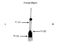 Frontal Force Diagram

- Sagittal
-
 Sagittal Force Diagram

References
edit- Baumgartner, T. J., & Baumgartner, D. R. (2009). U.S. Patent No. 7,597,671. Washington, DC: U.S. Patent and Trademark Office.
Manufacturing process
editLTT
- 1. Draw design and cut out template
- 2. Check that template fit the patient (make any necessary changes)
- 3. Use the template to cut out thermoplastic material
- 4. Submerge LTT into warm water and leave to heat
- 5. Position patient’s wrist and forearm and check angles
- 6. Remove LTT and slightly dry
- 7. Mould LTT to patients wrist and allow to set in correct position
- 8. Remove when set and neaten trimlines by submerging the edges in the water and trimming whilst warm.
- 9. Apply adhesive hook tape to volar surface at:
- 2nd to 5th digit proximal IPJ
- 1st digit IPJ
- radiocarpal joint and
- proximal trimline of orthosis.
- 10. Measure and cut the length needed for each of the 4 velcro loop straps (Round edges to look neat).
- 11. Apply velcro loop straps to the device.
- 12. Cut a piece of EVA wider than the 2nd-5th digit strap.
- 13. Make 2 incisions at each end to pass the velcro strap through.
- 14. Apply it the the digit strap to provide cushion against the phalanges.
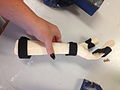
-
 Orthotic Template
 -
 LTT Template
 -
 LTT cooling after moulding


POP
- 1. Use the template to cut out POP slab
- 2. Position the patients arm at 30° wrist extension, 50° flexion of fingers and 40° abduction of the thumb.
- 3. Wet the POP slab and apply to the patients forearm.
- 4. Wait until set to remove and trim edges.
-
 POP device

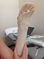
Critique of fit
edithttps://www.youtube.com/watch?v=DopEhhbRAMc
-
 Finished device
 -
 Fitted device (coronal view)
 -
 Fitted device (sagittal view)


Outcome measures
edit- There is no outcome measure chosen as the entire purpose of the orthosis was immobilisation of the wrist prior to surgical intervention as previous immobilisation had failed to reduce pain. This was chosen by the referring surgeon and was the initial and only requirement for the orthosis.
- If an outcome was to be measured it would be changes in pain after use of the orthoses. This could be measured on a scale. The scale chosen would be the numerical rating scale, more commonly referred to as the NRS. A study by Farrer, Young, LaMoreaux, Werth and Poole (2001) states that an 11-point pain intensity numerical rating scale (PI-NRS) is frequently used. This scale assesses the level of pain by indication of where the pain lies within a scale of 0-10, where 0 is no pain and 10 is the worst pain possible.
- This would have been chosen as it is a simple, easy to administer measure. Also, the test is easily repeatable. The reliability of the test is based on the subjects feelings and therefore brings doubt, and potential bias, when discussing the reliability. As it is open to influence by the subject the result may not always be a true reflection of the exact score. For example, if the patient is experiencing pain on the day the test is administered and is frustrated they may select 8/10 when the same pain on a good day is 6/10.
References
- Farrar, J. T., Young Jr, J. P., LaMoreaux, L., Werth, J. L., & Poole, R. M. (2001). Clinical importance of changes in chronic pain intensity measured on an 11-point numerical pain rating scale. Pain, 94(2), 149-158.
Direct Link to ULO Main Page
editComplete this for your client (using your clinical knowledge and judgement) for before and after the client receives the orthosis. Choose activies that you believe the client would have improved on and video your client undertaking these whilst wearing their orthoses. Outline your finding. Provide images of the completed Outcome measure on your wikipage.