Upper Limb Orthotics/Bennett Fracture with Subluxation
Describe your case study
editThe client is a 20 year old male who sustained a Bennett’s fracture of the left hand (dominant) from a fist fight that broke out outside a night club with other patrons. The fracture fragment is approximately 17% of the trapezium’s articular surface with significant displacement of 2mm. He is scheduled for closed reduction and percutaneous k-wire fixation and prescribed a low temperature thermoplastic wrist thumb spica orthosis to be worn for at least 4 weeks. The thumb should be immobilized in a mid-palmar and moderate adduction position. He is currently a 3rd year physiotherapy student who coaches basketball twice a week and plays once a week with the local club. As the injury has occurred in his dominant hand, it is advised that he does not participate in any basketball games, but may still coach, excessive movement may interrupt the reduction and prolong healing time. He will be unable to participate in all activities at university, although the orthosis allows movement at the inter-phalangeal joint of the thumb, this is not enough dexterity to grip a pencil or to perform any delicate handy work.
Written information
editEvidence
editAnatomy
The carpometacarpal (CMC) joint of the thumb is made up of the base of the first metacarpal and the trapezium bone. It is a biaxial saddle joint with smooth cartilage covering the articulating surfaces of each bone in addition it also has a loose capsule which allows for rotation, opposition and reposition with the other digits of the same hand (Hyde & Gengenbach, 2006). Hence it relies on surrounding ligament attachments for stabilization (Bettinger, Linscheid, Berger, Cooney III & An(1999). The thumb CMC joint axis rests in a pronated flexed position relative to the other fingers CMC joints, therefore it also behaves differently (Leversedge, 2008). The CMC joint abducts and adducts in a plane perpendicular to the palm and flex and extend in a parallel plane (Hyde & Gengenbach, 2006).
Pathology
When the base of the thumb breaks and dislocates (either partially or completely), this is known as a Bennett’s fracture. More specifically, it refers to an intra-articular fracture where the bone fragment is from the volar-ulnar edge of the first metacarpal base, but remains stabilized because of the anterior oblique ligament (Fufa & Goldfarb, 2012). In almost all Bennett fracture cases the muscular and ligament attachments remain intact, pulling the bone fragment and the metacarpal in different directions, which is the reason Bennett’s fractures are accompanied by dislocation or subluxation. One such attachment is the Abductor Pollicis Longus (APL) which pulls the thumb metacarpal dorsally, radially and proximally and dislocates the shaft (Jacobs, M., & Austin, N., 2013; Kozin, 2006). The Volar oblique ligament inserts into the base of the first metacarpal, opposes the action of the APL and holds the still attached volar bone fragment in anatomical position (Brownlie & Anderson, 2011). The long extensors of the thumb; the APL and the extensor pollicis brevis and longus as well as the thenar muscles all contribute to the dislocation that occurs with Bennett fractures(Leibovic, 1998) Dislocations or subluxations commonly occur with Bennett fractures due to the pull of surrounding ligaments. A qualified clinician needs to perform reduction on the joint. Closed reduction involves longitudinal traction on the thumb to pull the head of the first metacarpal distally while simultaneously pronating the thumb and applying pressure to push the metacarpal base medially to return it to anatomical position (Brownlie & Anderson, 2011 ;AO foundation). The difficulty associated with closed reduction is to correctly reduce the fracture (Brownlie & Anderson, 2011) and maintain the reduction sufficiently enough to allow healing in an anatomical position. (Ellis, 2013; Kjaer-Petersen, Langhoff & Andersen,1990; Oosterbos & De Boer, 1995), hence in many cases, practitioners choose to perform open reduction and/or internal fixation(Leibovic, 1998).
Mechanism of Injury
Bennett’s fracture is commonly caused by a forceful injury or any significant forces applied to the base of the first metacarpal, for example during sports games or falls. More precisely, it occurs when an adduction or axial force is applied to a partially flexed thumb (Hyde & Gengenbach, 2006; Carlsen & Moran, 2009)
Presentation
On presentation the proximal part of the first metacarpal will feel tender and appear swollen. The patient should feel weakness and difficulty when attempting to pincer grip or forming a fist and find they have a reduced range of motion in the thumb (Dial & Berg, 1972) Poorly treated Bennett’s fractures or fractures that have healed in a non-anatomical position have the potential to develop into arthritis in the future (Foster & Hastings, 1987). If left untreated, the thumb will show a reduced or lost range of movement in abduction and extension (Dial & Berg, 1972)
Orthotic treatment options
editOrthotic intervention can be used as management for Bennett fractures in a number of scenarios. Bennett’s fractures that have a relatively small fracture fragment without significant displacement and treated within 3 – 5 days of injury (Leibovic, 1998), can be managed with closed reduction and maintained with a short arm thumb spica plaster cast that holds the bones in correct alignment while it heals for 4 weeks (Leibovic, 1998). In these cases the wrist should be in neutral, the thumb CMC should be mid-palmar and in radial abduction (Jacobs & Austin, 2013). Older studies (Pollen, 1968) suggested that the thumb should be positioned in radial abduction, however a study of 12 hand cadavers with simulated Bennett’s fractures by Harvey and Bye suggest that the Bennett’s fracture should be immobolised in a moderate adduction (Harvey & Bye, 1976) because in this position the posterior oblique ligament is stretched which stablises the fragment. The spica cast should leave the inter-phalangeal joint of the thumb free, permitting the patient some thumb function while the fracture heals. (Fufa & Goldfarb, 2012) The downside of casts is the probability that skin ulcerations and hypoaesthesia may develop under cast pressure (Pollen, 1968). After removal of the cast, a orthoplast or low temperature thermoplastic thumb spica splint can be used while the patient gradually works to regain full thumb function (Leibovic, 1998). There is little evidence to suggest that you can replace the cast commonly used after closed reduction entirely with a thermoplastic orthosis. One reason for this gap in the existing literature may be that reduction occurs after putting on the plaster cast and while it is still wet, the mold is secured until the cast dries (Leibovic, 1998). There is the potential for a similar process to occur using custom made low temperature thermoplastics, moulding the plastic after it has been draped onto the patient, however there are currently no studies that either suggest or test this possibility. Conversely, on the market there exist an assortment of prefabricated wrist braces with accompanying thumb splint (physioroom, 2014) that claim to immobilize the wrist and thumb as well as a plaster cast. However there is little evidence to confirm the functionality of these braces besides the reviews left by customers on the site. Post surgery the patient is still required to wear a thumb spica cast or splint for 4 – 6 weeks to immobilize the joint and minimize any movement that may re-displace the joint. (Hyde & Gengenbach, 2006; Dial & Berg, 1972) Patients who are eligible for screw fixation and opt for it, are recommended to wear a thumb spica orthosis for 12 weeks post opt, because it provides added protection during activities (Hyde & Gengenbach, 2006).
Comparison of orthotic treatment options
editBennett’s fractures can be managed a number of ways; conservative treatment with closed reduction and splinting, closed reduction and percutaneous Kirschner wire (k-wire) fixation (Brownlie & Anderson, 2011) or open reduction and internal fixation (ORIF) (Fufa & Goldfarb, 2012). The goal of surgical intervention is to reduce the joint and improve congruency. Fractures that are more complicated with displacement greater than 1mm (Leibovic, 1998) require surgery to realign the bone fragments. In surgery an incision is made at the base of the thumb and screws, pins or plates are used to align the fracture in the correct position (Leibovic, 1998). The tools used to realign the bones are dependent on bone and fracture sizes and patient goals, for example; smaller fragments do not allow screw fixation so a 0.9mm k-wires would be used, but in athletes with bigger bones who want to return to game play as soon as possible, it is plausible to use 1.6mm screws which enables quicker return to function (Fufa & Goldfarb, 2012). When the fracture fragment is greater than 15 – 20% of the trapezium’s articular surface, it is recommended that closed reduction be accomplished using two kirschner wires (Hyde & Gengenbach, 2006). The procedure would require the wires to be placed percutaneously through the base of the thumb metacarpal and into the adjacent carpals or to the second metacarpal with the assistance of x-rays (Hyde & Gengenbach, 2006). Alternatively the two k-wires, can be inserted from the metacarpal to the fragment and the second pin from the metacarpal to the trapezium. (Fufa & Goldfarb, 2012; Hyde & Gengenbach, 2006). The consequence of using K-wire pin fixation is the potential for it to become infected (Wagner, 1950). If a fracture is significantly displaced and the bone fragment is large enough, a clinician can decide to perform an open reduction with a 1.5 to 2mm screw (Hyde & Gengenbach, 2006). The advantage of the screw fixation is that range of motion exercises can begin within 5 – 10 days instead of the standard 4 weeks required with closed reduction and fixation (Hyde & Gengenbach, 2006). However in these situations, instability is still a major concern, particularly in athletes who want to return their sport and place high stresses on their thumb (Fufa & Goldfarb, 2012). In these conditions, it is essential they wear a thumb spica brace (Hyde & Gengenbach, 2006).
Search Strategy
The databases Medline and Cinahl and search engine Google scholar were used to find the cited articles. The following terms were used: “Bennett fracture”, thumb, "spica splint", Ortho*,brace, treatment, management. Medline and google scholar yielded sufficient results when “bennett fracture” was searched, no limited to no relevant results were returned when “Bennett fracture” and “ortho*” or “brace” were searched together.
Functional Aims and Goals
editIn cases where the fracture fragment is larger than 15 – 20% of the trapezium’s articular surface and the first metacarpal has become significantly displaced closed reduction with k-wires or open reduction is necessary, however a wrist thumb spica is still required post surgery. The spica plays a minor role in reduction preservation of the CMC joint (most of which is maintained by the wire/screw fixation), it’s main goal is to provide protection of the joint and immobilising it in the correction position so that restriction to any thumb range of movement that may disrupt the reduction is minimised. In the case of the scenario where the fracture fragment is 17% of the trapezium's articular surface with a 2mm displacement, the wrist thumb spica will be fitted post internal fixation of the k-wires. It’s goal is to maintain the bones in correct alignment after reduction while the fracture heals for approximately 4 weeks. The functional aim of the wrist thumb spica is to hold the thumb in a moderate adduction because this allows the posterior oblique ligament to stablise the fracture fragment, while allowing full ROM of the inter-phalangeal joint of the thumb. The wrist thumb spica should also limit most wrist movements, as the fracture is at the base of the 1st metatarsal (articulating with the carpals) too much movement of the carpals can disrupt the reduction.
Design
edit
Wrist Thumb Spica Brace/Radial based thumb spica splint were traditionally made from plaster cast, but now there is more of a trend towards using low temperature thermoplastics due to the associated benefits. These include but are not limited to, improved intimacy of fit, adjustability and hygiene.
The proximal trim-line is 5 – 6cm distal to the cubital fossa so as to not limit any elbow range of motion. The distal trim-line is proximal to the mid palmar crease, so that hands and fingers still retain most functions. The thumb piece’s distal trim-line will be just proximal to the thumb IP joint, enough that it can still flex at this joint.
Wide Velcro straps will be used to distribute loading pressure as opposed to thin straps. Three strap will be utilized, proximal strap will be placed as close to the proximal trim-line as possible, here it ensures that when patient flexes elbow, the brace does not detach from the arm and disturb the integrity of the immobilization. The middle strap will wrap around the wrist and keep the wrist from lifting out of the brace and limit any flexion at the wrist. Finally the distal strap will extend from approximately mid-1st metatarsal, across the dorsum of the hand and onto the ulnar side. Proximal and middle hook Velcro piece are attached to the anterior surface of the orthosis via double sided tape and the straps wrap around the entire circumference. The distal Velcro hook piece uses the same tape on the ulnar side and the strap is heat gunned to the thumb site, as this is a smaller and more fragile strap.
Manufacturing process
edit
1. Position patient in a comfortable position on the plinth/working area.
2. Trace patient hand onto paper or fabric and mark the following landmarks
- Wrist joint on the ulnar and radial side
- Palmar crease (or mark the MCP joint of the 2nd and 5th digit and join the dots)
- Base of the 1st metacarpal
- Head of the 1st metacarpal
- IP joint of the thumb
- Approximately 2/3 of the distal forearm
3. Remove the patient’s hand and trace the template of the proposed base appropriately onto the hand tracing making sure to align to the hand
4. Cut out the template
5. Roughly fold over the patient’s hand to check that certain points of the template fit to landmarks of the hand, check the proximal length

6. Make any corrections if necessary
7. Trace the template onto the thermoplastic and cut out. Thermoplastic maybe need heating up to cut with normal scissors if shearers are not available.
8. Heat template again and fold the palmar crease edge
9. Position patient in ideal position
10. Rest the heated thermoplastic template for a few seconds on the plinth and dry it using a small towel, making sure to flip regularly or the template will adhere to any surface.

11. When the thermoplastic is at a reasonable temperature, approach the patient, let them know that the plastic is still warm and if it is too warm to alert us.
12. Apply the thermoplastic to the anterior side of the forearm and volar of the hand, pull the thumb piece between the thumb and index finger and wrap around the thumb just enough to reach the plastic on the side to form a loop. Lightly pinch the tip of the thumb and move in small circular motions to create a wider thumb hole
13. Ensure that distal trimline is below the mid palmar crease, if not adjust accordingly.
14. Smooth out the rest of the thermoplastic template over the anterior forearm, wrapping over to the posterior side. Apply gentle pressure at the wrist joint to ensure an intimate fit is achieved. Wrist should be in neutral position
15. Ensure that the thumb is still in the correct position relative to the hand, particular to this condition, the thumb should be moderately adducted
16. Let the plastic cool down sufficiently before removing
17. Radial trimline should not cover radial styloid and ulnar trim-line should not cover ulnar styloid, adjusting the width until the orthosis covers approximately the anterior half of the forearm
18. When cool check that all intended movements are still possible, if not adjust trimlines accordingly
19. Dip thumb piece into the hot water until malleable, and fold the distal trimline over so there are no jagged edges
20. Dip any protruding corners into the hot water for a few seconds, and round off the corners with scissors
21. Flare out proximal trimline slightly
22. Add Straps per design
Critique of fit
editClient is a 20 year old male who sustained a Bennett’s fracture of his right dominant hand from a fist fight that broke out outside a night club with other patrons. Client had apparently swung a punch but the other patron dodged, resulting in the client punching a brick wall. This caused the base of the 1st metatarsal or the thumb metatarsal to fracture. Clinical imaging revealed a fracture fragment approximately 17% of the trapezium’s articular surface and displaced 2mm.
Client coaches junior basketball and plays competitively once a week, and is currently a full time 3rd year physiotherapy student. The fracture in his dominant hand means he is unable to play basketball until it is healed or until the doctor believes it is safe to. He can still coach. 3rd year physiotherapy students receive a heavy work load, even though he is immobilized in a functional position (opposition still possible) it is difficult to grip a pen naturally or type. He’s ability to write at a certain pace is greatly limited as is the exercises he can perform as part of class work.
Traditionally he would have undergone surgery and casted in a forearm plaster of paris cast to maintain the reduction, however we have chosen to utilize more advanced materials; low temperature thermoplastics. The wrist thumb spica brace, keeps the thumb in the ideal position while limiting any wrist ranges of motion that may disrupt the k wire fixation. Originally a hand based thumb spica was prescribed, however on further investigation, it was determined that the base of the 1st metacarpal which articulates with the wrist/carpal bone; the trapezium may disrupt the fixation when moving. So a longer wrist immobilizing brace seemed more advisable. The device provides a relatively intimate fit, there are a few areas that have some leeway (fig. 1), which can be addressed by tightening the strap, but can also be achieved during molding of the device by wrapping the device tighter and holding on until the plastic has completely cooled down.
The proximal trim-line is ideal, sitting about 3 – 4 centimeters distal to the cubital fossa (fig. 2). However the distal trim-line would ideally be adjusted to be shorter or more proximal to the palmar crease. While the patient can still flex her hand at the palmar crease, it still appears slightly restricted (fig. 3).
-
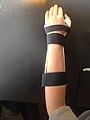 Fig.1 Relatively Intimate fit
 -
 Fig. 2 Approx 3 - 4 cm distal to the cubital fossa
 -
 Fig. 3 Slight restriction of hand flexion at palmar crease


When the thumb spica piece was molded, every effort was made for the patient to maintain the correct position when the thermoplastic was being applied; a neutral wrist, the thumb in opposition, still there was an issue with the thumb’s final position. Although the patient can oppose the thumb with each digit of the same hand, there is some extension at the mid palm to allow for it, the thumb is not as adducted towards the palm as it should be, the patient appears to strain the thumb to reach the other digits (fig. 4). We had tried to cast the thumb in a moderately adducted position as advised in the literature, however the degree we deemed appropriate during molding turned out to be less than that in the final product. In future the thumb spica would be cast with the thumb piece moderately adducted so that opposition is easier to perform. The thumb spica piece’s trim-line is ideal, ending just proximal to the Inter-phalangeal joint (fig. 5).
The fit of the rest of the device was satisfactory in fit and position, the lateral trimline clears the ulnar styloid (fig. 6) and the medial trim-line covers the medial styloid (fig. 7), which is not prominent, this is to further immobilize the thumb side. The lateral and medial trim-lines cover approximately half of the forearm circumference which is ideal (fig. 8).
-
 Fig. 4 Thumb spica not in enough flexion position
 -
 Fig. 5 Thumb spica distal trimline proximal to thumb IP joint
 -
 Fig. 6 Ulnar trimline under/clearing ulnar styloid
 -
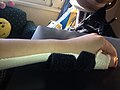 Fig. 7 Radial trimline covering radial styloid
 -
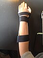 Fig. 8 Device covering necessary half of limb circumference


Outcome measures
editClick image for DASH forms, pre and post device.

Referral Letter
edit

References
editBettinger, P. C., Linscheid, R. L., Berger, R. A., Cooney III, W. P., & An, K. N. (1999). An anatomic study of the stabilizing ligaments of the trapezium and trapeziometacarpal joint. The Journal of hand surgery, 24(4), 786-798
Brownlie, C., & Anderson, D. (2011). Bennett Fracture Dislocation: Review and Management. Australian family physician, 40(6), 394.
Carlsen, B. T., & Moran, S. L. (2009). Thumb trauma: Bennett fractures, Rolando fractures, and ulnar collateral ligament injuries. The Journal of hand surgery, 34(5), 945-952.
Dial, W. B., & Berg, E. (1972). Bennett's fracture. The Hand, 4(3), 229-235.
Ellis, H. (2013). Edward Hallarran Bennett: Bennett's fracture of the base of the thumb. Journal of perioperative practice, 23(3), 59-60.
FOSTER, R. J., & HILL HASTINGS, I. I. (1987). Treatment of Bennett, Rolando, and vertical intraarticular trapezial fractures. Clinical orthopaedics and related research, 214, 121-129.
Fricker, R., Kastelec, M. & Nunez, F. (2008). Thumb – Bennett Fracture. AO Foundation. Retrieved from https://www2.aofoundation.org/wps/portal/surgery?bone=Hand&segment=Thumb&classification=76-Metacarpal,%20Bennett%20fracture&showPage=indication
Fufa, D. T., & Goldfarb, C. A. (2012). Fractures of the thumb and finger metacarpals in athletes. Hand clinics, 28(3), 379-388.
Harvey, F. J., & Bye, W. D. (1976). Bennett's fracture. The Hand, 8(1), 48-53.
Hyde, T. E., & Gengenbach, M. S. (Eds.). (2006). Conservative management of sports injuries. Jones & Bartlett Learning.
Jacobs, M. A., & Austin, N. M. (2013). Orthotic Intervention for the Hand and Upper Extremity: Splinting Principles and Process. Lippincott Williams & Wilkins.
Kjaer-Petersen, K., Langhoff, O., & Andersen, K. (1990). Bennett's fracture. The Journal of Hand Surgery: British & European Volume, 15(1), 58-61.
Kozin, S. H. (2006). Fractures and dislocations along the pediatric thumb ray.Hand clinics, 22(1), 19-29.
LEIBOVIC, S. J. (1998). Treatment of Bennett's and Roland0's Fractures. Techniques in hand & upper extremity surgery, 2(1), 36-46.
Leversedge, F. J. (2008). Anatomy and pathomechanics of the thumb. Hand clinics, 24(3), 219-229.
Physioroom. (2014). PhysioRoom.com Wrist Brace With Thumb Splint. Retrieved from http://www.physioroom.com/product/PhysioRoom.com_Wrist_Brace_With_Thumb_Splint/3079/38938.html
Pollen, A. G. (1968). The conservative treatment of Bennett’s fracture-subluxation of the thumb metacarpal. J Bone Joint Surg Br, 50(1), 91-101.
Wagner, C. J. (1950). Method of treatment of Bennett's fracture dislocation. The American Journal of Surgery, 80(2), 230-231.