Pharyngeal systems
Pharynges
editThe pharynx (plural: pharynges) is part of the neck and throat situated immediately posterior to (behind) the mouth and nasal cavity, and anatomical terms of location|cranial, or superior, to the oesophagus (esophagus), larynx, and vertebrate trachea|trachea. It is part of the digestive system and respiratory system of many organisms. It is an entranceway, a passageway (channel), a chamber, a concavity, and under special circumstances an outlet. It is also a connection and a separation.
Pharyngeal systems
editIn anatomy and zoology the pharynx is that part of the alimentary canal immediately behind the mouth involved in swallowing. In morphology it is a membrane-lined cavity also behind the nose (when present) which connects to the esophagus. Many organisms appear to have one. In the theory of the pharynx, one question is to determine its unique or novel purpose. For instance and however briefly, the pharynx serves as a container for extracellular fluid (ECF), specifically transcellular fluid (TCF), while breathing or swallowing is occurring. In some organisms, constriction or expansion of the body occurs associated with the pharynx.
A pharyngeal system may contain tissues and organs that are not found in the pharynx of an adult human. These tissues and organs may be considered as part of the pharynx due to anatomical or physiological inferences or deductions. As additional analytical techniques become available, their use may indicate whether those inferences or deductions are incorrect, perhaps incomplete, or factual. Phylogenetic analysis using molecular genetics and genomics is one such technique.
Gene expression in eukaryotes can often help to establish that particular organs occur at some developmental stage or have a special purpose. A mature mRNA template is one occurring in a cell where a gene is expressed as part of a particular cell's function. After addition to the cell reverse transcriptase scans the mature mRNA and synthesizes a sequence of DNA that complements the mRNA template.[1] This strand of DNA is complementary DNA (cDNA). Finding cDNA in Unigene, for example
- [1],
can help to establish that specific genes are expressed in tissues or organs such as the pharynx. Expression of these same or similar genes in organisms of questionable classification can help in their taxonomy.
Pharyngeal teeth
edit
Teeth occurring within the pharynx are usually referred to as pharyngeal teeth.
Pharyngeal teeth are teeth in the pharyngeal arch of the throat of cyprinids, Catostomidae (sucker)s, and a number of other fish species otherwise lacking teeth.[2]
Many popular aquarium fish such as goldfish and loaches have these structures. Members of the genus Botia such as clown loaches are known to make distinctive clicking sounds when they grind their pharyngeal teeth. Haemulidae (Grunts) are so called because of the sound they make when they grind them.[3] Molidae (Molas) are said to be able to produce sound by grinding their long, claw-like pharyngeal teeth.
The Chinese high fin banded shark (Myxocyprinus asiaticus) (family Catostomidae) has a single row of pharyngeal teeth with comb-like arrangements.[4] The Cape Fear shiner (family Cyprinidae) only has pharyngeal teeth, similar to the teeth of other omnivorous shiners.[5] The redear sunfish (family Centrarchidae) has thick pharyngeal teeth composed of hard, movable plates, which it uses to crush the exoskeletons of prey. The pharyngeal jaws of the moray eel (family Muraenidae) possess their own set of teeth. The dentary of the ghost knifefish species Sternarchogiton nattereri (family Ghost knifefish Apteronotidae) has upper and lower pharyngeal tooth plates bearing 9-11 and 7-9 teeth, respectively.
The mouth cone ("everted pharynx") of a possible new species of Meiopriapulus, a marine worm in the Priapulida, bears pharyngeal teeth.[6]
Fossils of the Yunnanozoon and Haikouella possess pharyngeal teeth.
Pharyngeal jaws
editPharyngeal jaws are a "second set" of jaws contained within an animal's mouth, or pharynx, distinct from the primary jaws. Or they are the only set of jaws an organism has.
Pharyngeal slits (pharyngeal stigmata)
editPharyngeal slits are used by organisms in feeding. The slits are separated by stiffening rods which allow a current of water to flow through. In fish the slits are modified for respiration. In tetrapods, they occur only in the embryo.
Ascidiacea
Pharyngeal gills
editSymplesiomorphy
Pharyngeal arches
editRecapitulation theory
Pharyngeal pouches
editOstracoderm
Pharyngeal pouch (embryology)
Pharyngeal muscles
edit- Stylopharyngeus muscle
- Salpingopharyngeus muscle
Constrictor muscle:
- Inferior pharyngeal constrictor muscle
- Middle pharyngeal constrictor muscle
- Superior pharyngeal constrictor muscle
Pharyngeal bones
editPharyngeal bones occur behind the gills in the oesophagus or gullet.
Platyzoa pharyngeal systems
editOne current scheme places the following traditional phyla in Platyzoa: Platyhelminthes, Gastrotricha, Gnathifera, Rotifera, Acanthocephala, Gnathostomulida, Micrognathozoa, and Cycliophora. The pharyngeal system within this superphyllum can include a cilial lining, the ability to evert the pharynx, secretion, powerful muscles, a jaw-like structure, bilateral symmetry, and lateral lamellae.
Gastrotricha myoepithelial pharynx
editThe myoepithelial pharynx of the Gastrotricha is an important distinguishing feature.[7] In the order Macrodasyida the pharynx has pharyngeal pores, while in the other order Chaetonotida, there is a pharyngeal plug.[7] The myoepithelial pharynx is also triradiate.[7]
Platyhelminthes pharynx
editSome of the turbellarians have a simple pharynx lined with cilia. Others can evert their pharynx to envelope prey. Some species of platyhelminths break up and soften food first by secreting enzymes in the gut or the pharynx (throat).[8]
Adult Digenea use a relatively large, muscular pharynx to ingest cells, cell fragments, mucus, body fluids or blood.
In some species of Monogenea the pharynx secretes enzymes that digest the host's skin, allowing the parasite to feed on blood and cellular debris.
Rotifera mastax
editRotifers have several ciliated tufts around the mouth that in motion resemble a wheel. These create a current that sweeps food into the mouth, where it is chewed up by a characteristic pharynx (called the mastax) containing a tiny, calcified, jaw-like structure called the trophi[9][10].
Acanthocephala
edit
Acanthocephalans are highly specialized parasites without mouth, pharynx and gut. Phylogenetic analysis of the 18S ribosomal gene has revealed that the Acanthocephala are most closely related to the rotifers, or may even belong in that phylum.
Gnathostomulida pharynx
editThe gnathostomulids are characterized by a specialized, muscular jaw, which they use to scrape smaller organisms off of the grains of sand that make up their anoxic seabed mud habitat.[11] This bilaterally symmetrical pharynx with its complex cuticular mouth parts make them appear closely related to rotifers and their allies, together making up the Gnathifera.
Micrognathozoa pharyngeal apparatus
editLimnognathia|Micrognathozoa show strong affinities with both Rotifera and Gnathostomulida (within the taxon Gnathifera), especially in the fine structure of the pharyngeal apparatus, where the jaw elements have cuticular rods with osmiophilic cores.[12] In Limnognathia maerski the main parts of the complicated jaw system have two lateral pharyngeal lamellae.[12]
Lophotrochozoa pharynx
editThe Lophophorata include the Bryozoa, Entoprocta, Phoronida, and Brachiopoda.
The lophophorates all feed by "impingement".[13]
Bryozoa pharynx
editThe Bryozoan gut is U-shaped, and consists of a pharynx which passes into the esophagus and cardia. The mouth opens into a relatively short, large, and muscular pharynx with a thick wall of tall columnar epithelial cells. The anterior region of the pharynx, into which the mouth opens, is ciliated. Food particles accumulate in the pharynx before being passed through the esophagus and cardia to the stomach.
In Gymnolaemata the feeding current is directed by the lophophore at the mouth where particles impinge and are sucked into the pharynx.[13] In Bryozoa|Phylactolaemata particles impinge in the trough of the lophophore and are transported to the pharynx by cilia.[13]
Phoronida pharynx
edit
In the Phoronids particles impinge on the frontal surface of the tentacles and are conveyed to the pharynx by tracts of cilia.[13]
Brachiopoda pharynx
editIn Brachiopods particles impinge on the frontal surface of the tentacles and are conveyed to the pharynx by tracts of cilia.[13]
Trochozoa pharynx
editThe Trochozoa include the Nemertea, Mollusca, Sipuncula, Echiura, and Annelida.
Nemertea pharynx
editThe pharynx in the Nemertea genus Cerebratulus has an ectodermal pharynx that is the first portion of the gut.[14]
Mollusca pharynx
editMollusks have pharyngeal glands (sugar glands) situated laterally to the pharynx.[15]
Annelida pharynx
edit
The Annelids, for example earthworms, have a pharynx. It is relatively strong due to the fact that the wall of the pharynx consists of epithelium with built up obliquely striated longitudinal and circular muscle layers between it and the nervous tissue. Earthworms, like many other annelids, suck their food. This sucking action is aided by the pharyngeal muscles.
In leeches, like the earthworm, the pharynx extends from the mouth to the esophagus which connects to the crop.
Ecdysozoa triradiate pharynx
editThe Ecdysozoa include the following phyla: Arthropoda, Onychophora, Tardigrada, Kinorhyncha, Priapulida, Loricifera, Nematoda and Nematomorpha.
Ecdysozoans have various features are found in the group, for instance, tardigrades, pycnogonids and roundworms have a triradiate pharynx.
Loricifera pharyngeal apparatus
editLoriciferans have unique adult features including the cuticular buccal tube and myoepithelial pharyngeal apparatus with placoids.[12] Annulation of the flexible buccal tube, telescoping of the mouth cone and three rows of placoids in the triradiated pharynx bulb are found only in Tardigrada and Loricifera.[12] The phylum Loricifera has several Cladistics|plesiomorphic characters, such as a Myoepithelial cells|myoepithelial pharyngeal bulb, comparable to the other Cycloneuralia, especially the Nematoda and Gastrotricha.[12] The parasitic Nematomorpha, whose adults lack the pharyngeal bulb, has a larva with an introvert similar to Scalidophora. The loriciferans have, therefore, been assigned to the Cephalorhyncha that includes Priapulida, Kinorhyncha and Nematomorpha.[12]
Tardigrada pharynx bulb
editThe pharynx is of a triradiate, muscular, sucking kind, armed with stylets. Annulation of the flexible buccal tube, telescoping of the mouth cone and three rows of placoids in the triradiated pharynx bulb are found only in Tardigrada and Loricifera.[12]
The Ecdysozoa-concept resolves the problem of the nematode-like pharynx and some data from 18S-rRNA and the HOX (homeobox) gene indicate a relation to roundworms.
Nematoda pharynx
editDeuterostomia pharyngeal systems
editEchinodermata pharyngeal systems
editAll five chordate characteristics are lacking in echinoderms.[16] Echinodermata have not as yet been reported to express the transcription factor Pax1/9.[16]
Echinoidea (sea urchins) pharynx
editIn images of the sea urchin (Strongylocentrous purpuratus) larva, produced with differential interference contrast optics, the pharynx is clearly visible and labelled.[17]
The pharynx in the sea urchin genuses Evechinus and Echinus is part of the alimentary canal, just after the mouth, and enclosed within Aristotle's lantern.[18] The lantern must be opened by dissection to observe the pharynx.[18] The pharynx leads into the narrow, much convoluted oesophagus.[18] The external covering of the pharynx is ciliated epithelium.[18]
Holothuroidea (sea cucumbers) pharyngeal bulb
editIn Holothuroidea|sea cucumbers an important feature is a calcareous ring that encircles the pharynx or throat.[19] The muscles that operate the oral tentacles are attached to this ring.[19]
Xenoturbellida pharyngeal elements
editAll five chordate characteristics (postanal tail, dorsal nerve cord, notochord, endostyle, and pharyngeal gill slits) are lacking in Xenoturbella.[16] Xenoturbella have not been reported to express the transcription factor Pax1/9.[16]
Hemichordata pharyngeal elements
editThree of the chordate characteristics are present in hemichordates: a ventral postanal tail, an endostyle, and pharyngeal gill slits.[16] Adult pharyngeal clefts have allied hemichordates with chordates.[20]
The hemichordate epibranchial ridge, like the chordate endostyle, is composed of specialized secretory cells, and these cells bind iodine. However, iodine binding occurs all throughout the pharynx.[16]
The hemichordate homolog of the gene NK2.1, which in chordates is expressed in the endostyle and the dorsal nervous system, is expressed in hemichordate larval neuronal structures and broadly in the pharyngeal endoderm, stomochord, and hindgut.[16] It appears that the endostyle function in chordates is accomplished broadly by the pharynx in hemichordates.[16]
The transcription factor Pax1/9 is expressed in the pharyngeal endoderm in hemichordates, cephalochordates, and in the vertebrate Gnathostomata|gnathostomes.[16]
Pharyngeal skeletal elements of hemichordates and cephalochordates are strikingly similar in appearance.[16]
Chordata pharyngeal system
editThe chordates have evolved a unique body plan within the deuterostomes and are considered to share five morphological characters: (1) a muscular postanal tail, (2) a notochord, (3) a dorsal neural tube, (4) an endostyle, and (5) Pharyngeal slit|pharyngeal gill slits.[16] Pharyngeal gills appear to function for feeding and oxygen absorption.[16] With the exception of tunicates, all deuterostomes with gills have structural elements associated with pharyngeal gills.[16] Studies of phylogeny have shown that it is likely the ancestral deuterostome was a worm that had pharyngeal gills.[16] Gills of vertebrates are developed in the walls of the pharynx along a series of gill slits opening to the exterior.
Urochordata pharyngeal elements
editPharyngeal gill slits are present in tunicates only in the tunicate tadpole larva.[16] The endostyle is an iodine-binding organ present in the tunicate pharynx.[16]
Cephalochordata pharyngeal elements
edit
Cephalochordates possess a pharynx.[16] The endostyle as an iodine-binding organ is also present in the cephalochordate pharynx.[16]
Cephalaspidomorphi pharyngeal system
editSee lamprey for a figure showing the muscular postanal tail.
Chondrichthyes pharynx
editChondrichthyes are jawed fishes with skeletons made of cartilage. Sharks, for example, have a pharynx.
Vertebrata pharyngeal system
editThe expression of the most anterior Hox gene, Hox1, is first seen in vertebrates in the second pharyngeal slit and the expression of Hox1 in hemichordates is seen at the level between the first and second gill slits.[16]
Neural crest cartilages are found in the vertebrate pharyngeal gills, and neural crest cells do take on the role of pharyngeal cartilage secretion.[16] The pharyngeal gills are supported by collagenous cartilage skeletons in the hemichordates, cephalochordates, and vertebrates.[16]
Osteichthyes pharynx
editThe Osteichthyes are bony fish that primarily breathe though gills and they have a pharynx.
Amphibia pharynx
editFrogs are amphibians that have gills during their tadpole stage and lungs as adults. Their digestive system begins with a mouth, continues with a pharynx, oesophagus, stomach, small intestine, and large intestine.
Reptilia pharynx
editAves pharynx
editBirds have a throat. Many birds possess a muscular pouch along the oesophagus called a crop.
Mammalia pharynx
editHuman pharynx
editA pharynx is found as part of the neck and throat. A mouth and nasal cavity occur anteriorly. As part of the neck, it is superior to the oesophagus, larynx, and trachea. Viewed from outside the body, there is a constriction and concavity associated with the pharynx. The pharynx does connect the mouth to the oesophagus and the trachea. A membrane serves as a lining.
Morphology of the pharynx in humans
editHumans follow the unique body plan of the chordates and have the five morphological characters: (1) a muscular postanal tail (vestigial), (2) a notochord (vertebral column), (3) a dorsal neural tube (spinal cord), (4) an endostylic thyroid gland, and (5) pharyngeal gill slits (during early development). With the exception of the tail, each character has a portion in the neck near to or as part of the pharynx.
All species of the genus Homo (genus)|Homo except Homo sapiens (H. sapiens) (modern humans) are Extinction|extinct. All the subspecies of H. sapiens are extinct except Homo sapiens sapiens (H. s. sapiens). Although many (and probably all) of the morphological characteristics observable in H. s. sapiens also occurred in other members of the genus, for gross observational consideration only specimens of H. s. sapiens are available.
Connection
edit


From the side view exterior as suggested by the throat diagram, the characteristic location of the pharynx appears to be primarily within the lower part of the head and upper part of the neck. By looking at the photograph of the side view of an individual human female and comparing it with the throat diagram, it may be concluded that only the lowest portion of the pharynx is actually in the neck.
Viewed from the front only, as seen in the closeup front view of an individual human male when compared to the throat diagram, it may be concluded that the pharynx is not part of the throat.
To demonstrate that these two photographs are not unique, consider the collection of front view photographs.
From visual inspection exteriorly of humans on a macroscopic scale, it may be concluded that the pharynx does not serve as a connection between the head and thorax.
Concavity
editAs can be seen in the photographs referred to in the section on connection, the concavity is associated with the exterior features of the neck and throat of a human. But because of the location of the pharynx in humans, it is unlikely that it functions as part of the exterior concavity between human head and thorax. The pharynx's close proximity to the division between head and neck suggests that it may play a role in the separation between head and neck.
Separation
editBecause of the presence of a neck and throat, a human's head is separated from its thorax or torso. While not wholly contained within either the neck or throat, the position of the pharynx at the division between head and neck or throat, and location nearly entirely within the head may be related more with the onset of concavity and the connection between head and throat than the connectivity of the head to the thorax.
Entranceway
edit
The mouth (oral cavity) or buccal cavity is the Head and neck anatomy|entranceway into the digestive system.


As the drawing of the open human mouth shows, the posterior wall of the pharynx is part of an entranceway. The photograph of an open human mouth also contains a partial image of the pharynx behind the uvula.
In addition to the mouth, humans have another entranceway in the nose.
Of the many entranceways into the human body, the nasal part of the pharynx always remains open with respect to entry through the nose. However, when swallowing the nasal portion of the pharynx is sealed off from the mouth.
When breathing through either the nose or mouth, the pharynx is open; however, breathing through both openings simultaneously may not be possible. Breathing through the nose while the mouth is open occurs after the back of the tongue presses upward to close off the pharynx from the mouth.
It is clearly visible that the pharynx is part of an entranceway into a human body.
The abdominal portion of a human also has several entranceways into the body. However, no portion of the pharynx is readily visible from any abdominal entranceway. As part of the digestive system, some of these entranceways serve primarily egestive functions. See, for example, defecation and urination.
From its location in the head end of a human, it appears that the pharynx is involved more with ingestion than egestion.
Passageway
edit
Although passageways or Channel (geography)|channels are often perceived two-dimensionally, a channel is also a Tubing (material)|tubular Passage (architecture)|passage or Duct (anatomy)|duct for fluid. For the pharynx, while humans breathe, Earth's atmosphere|air passes from the mouth into the lungs.
When a human Swallowing|swallows chewed solids or drunk liquids, the tongue retracts in a posterior direction to force the bolus into the pharynx. Peristalsis advances the bolus from the pharynx to the esophagus. In this way, the pharynx also serves in passage as a passageway.
Chambers
edit
A chamber is any distinquishable space within a structure or an enclosed space. In the previous section about passageways, the diagram of conducting passages points out a space that appears to be distinguishable from the nasal cavity and the larynx.
The esophagus is continuous with the laryngeal part of the pharynx. There appear to be no valves between the esophagus and the pharynx. But during swallowing, the epiglottis folds to a more horizontal position. It serves as a valve between the trachea and the pharynx. When breathing there is a constriction of the junction below the epiglottis between the pharynx and the esophagus.
Breathing through the nose while the mouth is open occurs apparently after the mouth is closed-off from the pharynx. In addition, as seen in the throat diagram of the connection section above there is a visible reduction in cavity width of the nasal cavity between it and the pharynx.
It seems readily apparent that the pharynx is a chamber.
Outlets
editPart of human breathing is Exhalation|exhaling. Air is moved out of the lungs, through the airways, to the external environment. For humans the airway begins at the mouth or nose and accesses the trachea via the pharynx. Air (or water vapor) can be seen exiting a human mouth on a very cold day. It can also be felt as it rushes out the nose or mouth.
There are outlets from the human body that function far more often as outlets than inlets, see the entranceway section above, and pelvic outlet. Regarding breathing the pharynx serves as part of an outlet.
As part of the digestion system, the pharynx is included or serves as a passageway during vomiting|emesis. With emesis often involving the mouth of which the pharynx is a part, it is reasonable to assume that the pharynx serves as an occasional outlet.
Anatomy of the human pharynx
edit

Arteries: pharyngeal branches of ascending pharyngeal artery, ascending palatine artery (ascending palatine), descending palatine artery (descending palatine), pharyngeal branches of inferior thyroid artery (pharyngeal branches of inferior thyroid).
The human pharynx is conventionally divided into three sections:
Nasopharynx
editThe nasopharynx lies behind the nasal cavity.
Postero-superiorly this extends from the level of the junction of the hard and soft palates to the base of skull, laterally to include the fossa of Rosenmüller.
The inferior wall consists of the superior surface of the soft palate. Like the nasal passages it is lined with pseudostratified ciliated columnar epithelium.
Oropharynx
editThe oropharynx lies behind the oral cavity and is lined by stratified squamous epithelium. The anterior wall consists of the base of the tongue and the vallecula; the lateral wall is made up of the tonsil, tonsillar fossa, and tonsillar (faucial) pillars; the superior wall consists of the inferior surface of the soft palate and the uvula.
Laryngopharynx
editThe laryngopharynx, also known as the hypopharynx, roughly corresponds to the levels between C4 to C6, it includes the pharyngo-esophageal junction (postcricoid area), the piriform sinus, and the posterior pharyngeal wall.
Like the oropharynx above it the hypopharynx serves as a passageway for food and air and is lined with a stratified squamous epithelium.
It lies inferior to the upright epiglottis and extends to the larynx, where the respiratory and digestive pathways diverge.
At that point, the laryngopharynx is continuous with the esophagus posteriorly. The esophagus conducts food and fluids to the stomach; air enters the larynx anteriorly. During swallowing, food has the "right of way", and air passage temporarily stops. The upper esophageal sphincter opens during swallowing so the bolus can pass into the esophagus. See also the pharyngeal constrictor.
Parapharyngeal space (PPS)
editThe PPS is in the shape of an inverted triangular pyramid with concave faces, going from the base of the skull to an apex (the greater cornu) of the hyoid bone, up to the petrotympanic fissure of the temporal bone (petrous part of the temporal bone). The back wall is delineated by the pharyngeal aponeurosis and the prevertebral muscles of the cervical vertebrae C1, C2 and C3. It is delimited medially by the pharyngobasilar fascia and the superior pharyngeal constrictor muscle, and laterally by the Ramus of the mandible|ascending branch of the jaw (by the medial pterygoid muscle), palatine aponeurosis and the Submandibular gland|submaxillary gland.[21] The parapharyngeal space curves around the lateral side of the pharyngeal mucosal space and is bounded from below by the muscles arising from the styloid process.
The PPS is divided by an Aponeuroses|aponeurotic sheath originating in the apophysis of the styloid process into three compartments: the prestyloid, occupied mainly by the deep lobe of the parotid gland, the post styloid, containing the internal carotid artery (carotid space), internal jugular vein, cervical sympathetic chain and the last four pairs of cranial nerves, and the retropharyngeal space, which contains a lymph node present mainly during childhood.
Physiology of the human pharynx
editEpiglottis
editBecause both food and Earth's atmosphere|air pass through the pharynx, a flap of connective tissue called the epiglottis, part of the oropharynx, closes over the trachea when food is swallowed to prevent choking or Pulmonary aspiration|aspiration. It is normally pointed upward during breathing with its underside functioning as part of the pharynx, but during swallowing, elevation of the hyoid bone draws the larynx upward; as a result, the epiglottis folds down to a more horizontal position, with its upper side now functioning as part of the pharynx. In this manner it prevents food from going into the trachea and instead directs it to the esophagus, which is more posterior.
The epiglottis is one of nine cartilaginous structures that make up the larynx (voice box). Yet while breathing it lies completely within the pharynx. When swallowing it serves as part of the anterior of the larynx.
Pharyngeal consonant manner of articulation
editSome languages, such as Arabic, use the pharynx to articulate some sounds.
Pharyngeals are known primarily from two areas of the world: in North-Africa/Mideast (in the Semitic languages: Semitic, Berber languages: Berber, Cushitic languages: Cushitic, Northwest Caucasian languages: Circassian, and Northeast Caucasian languages: Dagestanian families) and in British Columbia (in the Wakashan languages: Wakashan and Salishan languages: Salish families). There are scattered reports of pharyngeals elsewhere, such as in the Nilo-Saharan Tama language and in Nenets language: Nenets in Nenetsia: Northern Russia. In Finnish language: Finnish, a weak pharyngeal fricative is the realization of /h/ next to the vowel /a/, but since this is mere allophony, it is transcribed as /h/. According to the Laryngeal theory#Pronunciation (laryngeal theory), the Proto-Indo-European language might also have contained pharyngeal consonants.
Note that reported pharyngeals frequently turn out to be Epiglottal consonant|epiglottals. Such was the case for Dahalo language: Dahalo and northern Haida language: Haida, for example, and is likely to be true for many if not most of the others. This is perhaps because 'epiglottal' was only recently recognized as a distinct place of articulation, rather than a variant of 'pharyngeal'. The only language known to have contrastive pharyngeals and epiglottals is Aghul languages|Agul, a Northeast Caucasian languages: Lezgian language of Dagestan.
Secretion within the pharynx
editRespiratory tract secretions consist of mucus, surfactant (separating the mucous and periciliary layers), and periciliary fluid (between mucous layer and the epithelium).[22] Airway mucus (a nonhomogeneous, adhesive, viscoelastic gel composed of water, carbohydrates, proteins, and lipids) is the secretory product of goblet cells and the submucosal glands.[22]
Mucus is a slippery secretion produced by, and covering, mucous membranes. The average human body produces about a liter of mucus per day.[23] It is a viscous colloid, containing antiseptic enzymes (such as lysozyme) and immunoglobulins, that serves to protect epithelial cells in the respiratory, gastrointestinal, and auditory systems of humans. Mucus also contains mucins, produced by goblet cells in the mucous membranes and submucosal glands, and inorganic salts suspended in water.
Mucus is a gel, primarily a 3-dimensional tangled polymer network of mucins.[22] Mucin macromolecules are 70-80% carbohydrate, 20% protein, and 1-2% sulfate bound to oligosaccharide side chains.[22] The protein backbone of each mucin is encoded by a mucin gene (MUC gene), at least 8 of which are expressed in the respiratory tract.[22] MUC5AC and MUC5B are the two principal gel-forming mucins secreted in the airway.[22]
Gene ID: 4584 is Mucin 3A (MUC3A).[24] cDNA source: pharynx.[25]
Gene ID: 4585 is Mucin 4 (MUC4).[26] Isoform a cDNA source: pharynx.[27]
Gene ID: 727897 is Mucin 5B (MUC5B).[28] cDNA source: pharynx.[29]
Gene ID: 10071 is Mucin 12 (MUC12).[30] cDNA source: pharynx.[31]
Gene ID: 94024: is Mucin 16 (MUC16).[32] cDNA source: pharynx.[33]
Gene ID: 4162 is Mucin 18 (MCAM).[34]. cDNA source: pharynx.[35]
Gene ID: 283463 is Mucin 19 (MUC19).[36]. The gel-like properties of mucus, which covers the epithelial surface of various tissues, are generally attributed to the gel-forming mucins such as MUC19.[36]
Gene ID: 200958 is Mucin 20 (MUC20).[37]. Isoform L cDNA source: pharynx.[38]
Gene ID: 394263 is Mucin 21 (MUC21).[39] cDNA source: pharynx.[40]
In addition to secretion, mucus is transported from the lower respiratory tract into the pharynx by air flow and mucociliary clearance.[22]
Immunoglobulin-A[41]
Secretions of the oropharynx
editAspiration of oropharyngeal or gastric secretion can contribute to lung abscess.
The pharynx as a container
editAn adult human is usually breathing and often swallowing. A human can hold its breath and refrain from swallowing for short periods. During these periods or when breathing or swallowing, the pharynx serves as a container for ECF, TCF, and ingestives. To prevent something from entering the throat except as part of normal swallowing, there is the pharyngeal reflex.
Chiefly in zoology, a receptacle is an organ or structure that receives a secretion. A container is often thought of as having sides, a bottom, and a top (sometimes serving as a lid). While the pharynx does not have a lid, it does have the nasal cavity on top of it. And, when the epiglottis and upper esophageal sphincter are closed, the pharynx has a bottom.
The rhinopharynx (nasopharynx) is the nasal part of the pharynx, lying above the level of the soft palate (upper surface of the soft palate).[42][43]
Cell biology of the human pharynx
editIn the nasopharyngeal tonsils (adenoids), lymphoepithelium (consisting of ciliary epithelium, non-ciliary epithelial cells, M cells, goblet cells, and many intraepithelial lymphoid cells) covers the nasopharyngeal lymphoid tissue.[44]
Goblet cell
editGoblet cells and ciliated cells are present throughout the rhinopharynx and in the upper half of the pharynx in human fetuses and prematures.[45]
M cell (microfold cell)
editMicrofold cells have broad, irregular cytoplasm-containing microvilli on their surface and small vesicles in their cytoplasm.[44]
Illnesses of the human pharynx
editIn persons with hayfever, oral allergy syndrome and related allergies, the pharynx is often a reaction site to allergens, with common symptoms including burning and itching.
Carcinoma
editNeck squamous cell carcinoma, an oropharyngeal cancer, occurs in humans.[46]
Inflammation
editPharyngitis is an inflammation of the throat or pharynx.[47] In most cases it is painful, and thus is often referred to as a sore throat. Inflammation of the tonsils (tonsillitis) and/or larynx (laryngitis) may occur simultaneously, which can make eating difficult or painful.
Most cases are caused by virus|viral infections (40%-60%), with the remainder caused by bacterial infections, fungus|fungal infections, or irritants such as pollutants or chemical substances.[48]
Treatment of viral causes are mainly symptomatic while bacterial or fungal causes may be amenable to antibiotics and antifungals respectively.
Hyperplasia
editSmoking-induced nasopharyngeal lymphoid hyperplasia occurs in heavy smokers.[49]
Additional images
edit-
 Conducting passages.
 -
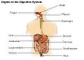 Organs of the digestive system.
 -
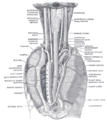
 The entrance to the larynx, viewed from behind.
 -
 Sagittal section of nose mouth, pharynx, and larynx.
 -
 The position and relation of the esophagus in the cervical region and in the posterior mediastinum. Seen from behind.


See also
edit- Adenoid
- Eustachian tube
- Hyoid
- Keratosis pharyngis
- Larynx
- Tonsil
- Uvula
References
edit- ↑ "cDNA - Definitions from Dictionary.com". dictionary.reference.com. Retrieved 2008-04-26.
- ↑ "Suckers Catostomidae", Iowa Department of Natural Resources
- ↑ Rocha, Luiz A.; Lindeman, Kenyon C.; Rocha, Claudia R.; Lessios, H.A. (2008). "Historical biogeography and speciation in the reef fish genus Haemulon (Teleostei: Haemulidae)". Molecular Phylogenetics and Evolution 48 (3): 918–928. doi:10.1016/j.ympev.2008.05.024. http://stri.si.edu/sites/publications/PDFs/Rocha_etal_Lessios_2008_Haemulon_phylogenetics.pdf.
- ↑ Myxocyprinus asiaticus, Chinese sucker fish, Filamn.ifm-Geomar.de, 2006, retrieved on: August 21, 2007
- ↑ Groves, John. "Cape Fear Shiner Conservation & Research". North Carolina Zoo. Archived from the original on 2007-10-21. Retrieved 2007-12-09.
- ↑ "First Molecular Data on the Phylum Loricifera – An Investigation into the Phylogeny of Ecdysozoa with Emphasis on the Positions of Loricifera and Priapulida". Zoological Science 23 (11): 943–54. Nov 2006. doi:10.2108/zsj.23.943. PMID 17189906.
- ↑ 7.0 7.1 7.2 Hochberg R, Litvaitis MK (Apr 2000). "Phylogeny of Gastrotricha: a morphology-based framework of gastrotrich relationships". Biol Bull. 198 (2): 299-304. PMID 10786949.
- ↑ Walker J.C.; Anderson D.T. (2001). "The Platyhelminthes". In Anderson, D.T.. Invertebrate Zoology. Oxford University Press. pp. 58–80. ISBN 0195513681.
- ↑ Klusemann J; Kleinow W; Peters W (Apr 1990). "The hard parts (trophi) of the rotifer mastax do contain chitin: evidence from studies on Brachionus plicatilis". Histochemistry. 94 (3): 277-83. PMID 2401635.
- ↑ Fontaneto D, Melone G (2006). "Postembryonic development of hard jaws (trophi) in a species belonging to the Brachionus plicatilis complex (Rotifera, Monogononta): a morphometric analysis". Microsc Res Tech. 69 (4): 296-301. PMID 16586490.
- ↑ Barnes, R.F.K. et al (2001). The Invertebrates: A Synthesis. Oxford: Blackwell Science.
- ↑ 12.0 12.1 12.2 12.3 12.4 12.5 12.6 Kristensen RM (2002). "An Introduction to Loricifera, Cycliophora, and Micrognathozoa". Integr Comp Biol. 42 (3): 641-51. doi:10.1093/icb/42.3.641.
- ↑ 13.0 13.1 13.2 13.3 13.4 Bullivant JS (1968). "THE METHOD OF FEEDING OF LOPHOPHORATES (BRYOZOA, PHORONIDA, BRACHIOPODA)". NZ J Mar Freshwat Res. 2: 135-46. http://www.royalsociety.org.nz/Site/publish/Journals/nzjmfr/1968/10.aspx.
- ↑ "Cerebratulus Ribbonworm Invertebrate Anatomy OnLine".
- ↑ Sturm, Charles F; Timothy A. Pearce; Ángel Valdés (2006). The Mollusks: A Guide to Their Study, Collection, and Preservation. Universal-Publishers. pp. 220, 445. ISBN 9781581129304.
- ↑ 16.00 16.01 16.02 16.03 16.04 16.05 16.06 16.07 16.08 16.09 16.10 16.11 16.12 16.13 16.14 16.15 16.16 16.17 16.18 16.19 16.20 Rychel AL; Smith SE; Shimamoto HT; Swalla BJ (Mar 2006). "Evolution and development of the chordates: collagen and pharyngeal cartilage". Mol Biol Evol. 23 (3): 541–9. doi:10.1093/molbev/msj055. PMID 16280542. http://mbe.oxfordjournals.org/cgi/content/full/23/3/541.
- ↑ Arenas-Mena C; Cameron AR; Davidson EH (Nov 2000). "Spatial expression of Hox cluster genes in the ontogeny of a sea urchin". Development. 127 (21): 4631–43. PMID 11023866. http://dev.biologists.org/cgi/reprint/127/21/4631.
- ↑ 18.0 18.1 18.2 18.3 McRae A (May 1959). "Evechinus chloroticus (Val.), an Endemic New Zealand Echinoid". Trans Proc Roy Soc New Zealand 1868-1961. 86 (3 & 4): 205–67. http://rsnz.natlib.govt.nz/volume/rsnz_86/rsnz_86_03_002080.html.
- ↑ 19.0 19.1 "Holothuroidea Sea cucumbers".
- ↑ Blair JE, Hedges SB (Nov 2005). "Molecular Phylogeny and Divergence Times of Deuterostome Animals". Mol Biol Evol. 22 (11): 2275-84. doi:10.1093/molbev/msi225. PMID 16049193. http://mbe.oxfordjournals.org/cgi/content/full/22/11/2275.
- ↑ Fernández Ferro M; Fernández Sanromán J; Costas López A; Sandoval Gutiérrez J; López de Sánchez A (Jan 2008). "Surgical treatment of benign parapharyngeal space tumours. Presentation of two clinical cases and revision of the literature". Med Oral Patol Oral Cir Bucal. 13 (1): E61-4. PMID 18167484.
- ↑ 22.0 22.1 22.2 22.3 22.4 22.5 22.6 Rubin BK (July 2002). "Physiology of airway mucus clearance". Respir Care. 47 (7): 761-8. PMID 12088546.
- ↑ "What's a Booger?".
- ↑ "Entrez Gene: MUC3A mucin 3A, cell surface associated".
- ↑ "Unigene: Mucin (MUC3) (MUC3A)".
- ↑ "Entrez Gene: MUC4 mucin 4, cell surface associated".
- ↑ "Unigene: Tracheo-bronchial mucin (MUC4) (MUC4)".
- ↑ "Entrez Gene: MUC5B mucin 5B, oligomeric mucus/gel-forming".
- ↑ "Unigene: Gallbladder mucin MUC5B (MUC5B)".
- ↑ "Entrez Gene: MUC12 mucin 12, cell surface associated".
- ↑ "Unigene: Mucin 11 (MUC11) (MUC12)".
- ↑ "Entrez Gene: MUC16 mucin 16, cell surface associated".
- ↑ "Unigene: Transcribed locus, strongly similar to NP_078966.2 mucin 16 [Homo sapiens]".
- ↑ "Entrez Gene: MCAM melanoma cell adhesion molecule".
- ↑ "Unigene: MUC18 glycoprotein (MCAM)".
- ↑ 36.0 36.1 "Entrez Gene: MUC19 mucin 19, oligomeric".
- ↑ "Entrez Gene: MUC20 mucin 20, cell surface associated".
- ↑ "Unigene: Mucin 20, cell surface associated (MUC20), transcript variant L, mRNA (MUC20)".
- ↑ "Entrez Gene: MUC21 mucin 21, cell surface associated".
- ↑ "Unigene: CDNA FLJ50027 complete cds, weakly similar to Dentin sialophosphoprotein precursor (MUC21)".
- ↑ Yagame M; Tomino Y; Eguchi K; Miyazaki M; Miura M; Suga T; Endoh M; Nomoto Y et al. (Sep 1987). "Correlation between the pharyngeal secretion of IgA, secretory-IgA and free secretory component and the upper respiratory tract infections in patients with IgA nephropathy". Nippon Jinzo Gakkai Shi. 29 (9): 1101-5. PMID 3694888.
- ↑ "Rhinopharynx".
- ↑ "rhinopharynx".
- ↑ 44.0 44.1 {{ cite journal |author=Fujimura Y |title=Evidence of M cells as portals of entry for antigens in the nasopharyngeal lymphoid tissue of humans |journal=Virchows Arch. |volume=436 | issue=6 |pages=560-6 |date=Jun 2000|pmid=10917169 ||
- ↑ Tos M (Aug 1975). "Goblet cells in the developing rhinopharynx and pharynx". Arch Otorhinolaryngol. 209 (4): 315-24. PMID 1243635.
- ↑ Almadori G; Bussu F; Galli J; Limongelli A; Persichilli S; Zappacosta B; Minucci A; Paludetti G et al. (July 2007). "Salivary glutathione and uric acid levels in patients with head and neck squamous cell carcinoma". Head Neck. 29 (7): 648-54. PMID 17274058.
- ↑ Dorlands Dictionary, volume six, pharyngitis
- ↑ "eMedicine - Pharyngitis : Article by John R Acerra".
- ↑ Finkelstein Y; Malik Z; Kopolovic J; Bernheim J; Djaldetti M; Ophir D (Dec 1997). "Characterization of smoking-induced nasopharyngeal lymphoid hyperplasia". Laryngoscope. 107 (12 Pt 1): 1635-42. PMID 9396678.
External links
edit- Schematic representations of the two orders within Gastrotricha showing the difference in pharynges.
- Some beautiful images of Rotifer trophi.
- A good example of the jaws or trophi of the rotifer Brachionus.
- - great transmission electron micrograph of the Limnognathia jaw showing the pharyngeal lamella.
- contains microphotographs of Phoronida including one of the pharynx.
- Internal anatomy of an earthworm (mouth to gizzard) showing the pharynx - lateral section.
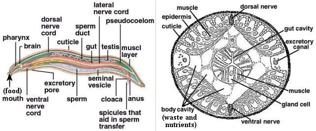
- Diagram of a roundworm shows the location of the pharynx.
- Strongylocentrous purpuratus larva image clearly shows the pharynx labelled.
- The main internal anatomical features of the sea cucumber Dendrochirotida include a pharyngeal bulb.
- An excellent diagram of the internal structure of a tunicate adult contains a well-labelled pharynx with gill slits (also labelled). The image following of a tunicate larvae has the pharynx with gill slits labelled.
- The internal structure diagram of a lancet clearly shows and labels the gill slits in the pharynx. The following image of the anterior anatomy of Amphioxus exhibits the gill slits and gill bars.
- Dissection of a lamprey with the pharynx, gills, spinal cord, and notochord labelled.
- Diagrams the invertebrate anatomy of the lamprey and labels the endostyle.
- A diagram displays the internal anatomy of a shark, with organs labelled including the pharynx and inner spiracle openings.
- Diagram of the vertical lateral cross section of the head and thorax of a vertebrate fish with the pharynx labelled.
- Diagram of the internal organs of a frog including a labelled pharynx.
- Verticle lateral cross section of a cat shows the nasopharynx.
- Pharynx, Stedman's Online Medical Dictionary at Lippincott Williams and Wilkins
- Human Anatomy and Physiology Elaine N. Marieb and Katja Hoehn, Seventh Edition.
- Template:SUNYAnatomyLabs
- TNM Classification of Malignant Tumours Sobin LH & Wittekind Ch (eds)Sixth edition UICC 2002 ISBN 0-471-22288-7
{{Gene project}}
Learn more about Pharyngeal systems |
Learn more about Pharynx |