File:Brain - Lobes.png
No higher resolution available.
Brain_-_Lobes.png (701 × 487 pixels, file size: 360 KB, MIME type: image/png)
| This is a file from the Wikimedia Commons. The description on its description page there is shown below.
Commons is a freely licensed media file repository. You can help. |
| Description |
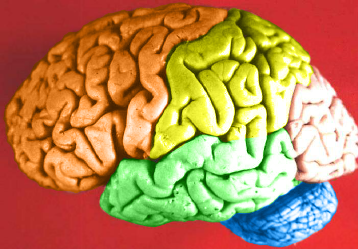
Human brain lateral view - Lobes
|
| Date | (UTC) |
| Source | Human_brain_lateral_view_description.JPG |
| Author | Dep't. of Cellular Biology & Anatomy, Louisiana State University Health Sciences Center Shreveport |
| Permission (Reusing this file) |
CC-BY |
| Other versions |
| This is a retouched picture, which means that it has been digitally altered from its original version. Modifications: Hemispheres in color.. The original can be viewed here: Human brain lateral view description.JPG:
|
Licensing
I, the copyright holder of this work, hereby publish it under the following license:
This file is licensed under the Creative Commons Attribution 2.5 Generic license.
- You are free:
- to share – to copy, distribute and transmit the work
- to remix – to adapt the work
- Under the following conditions:
- attribution – You must give appropriate credit, provide a link to the license, and indicate if changes were made. You may do so in any reasonable manner, but not in any way that suggests the licensor endorses you or your use.
The following refers to the original source file, not this derivative version.
This file, which was originally posted to
https://web.archive.org/web/20110514023714/http://www.healcentral.org/healapp/showMetadata?metadataId=40566, was reviewed on 1 November 2013 by reviewer Avenue, who confirmed that it was available there under the stated license on that date.
|
Original upload log
This image is a derivative work of the following images:
- File:Human_brain_lateral_view_description.JPG licensed with Cc-by-2.5
- 2006-06-20T13:58:22Z Patho 701x487 (50176 Bytes) {{Information| |Description='''Human brain lateral view - Lobes''' # Lobus frontalis # Lobus parietalis # Lobus temporalis # Lobus occipitalis # Sulcus lateralis # Sulcus centralis # Sulcus parietooccipitalis # Incisura preo
- 2006-06-20T13:54:13Z Patho 701x487 (49891 Bytes) Auf eine alte Version zurückgesetzt
- 2006-06-20T13:51:38Z Patho 701x487 (50074 Bytes) {{Information| |Description='''Human brain lateral view - Lobes''' # Lobus frontalis # Lobus parietalis # Lobus temporalis # Lobus occipitalis # Sulcus lateralis # Sulcus centralis # Sulcus parietooccipitalis # Incisura preo
- 2006-06-20T13:28:44Z Patho 701x487 (49891 Bytes) {{Information| |Description='''Human brain lateral view''' # Lobus frontalis # Lobus parietalis # Lobus temporalis # Lobus occipitalis # sulcus lateralis # Sulcis centralis # Sulcus parietooccipitalis # Incisura preoccipital
Uploaded with derivativeFX
File history
Click on a date/time to view the file as it appeared at that time.
| Date/Time | Thumbnail | Dimensions | User | Comment | |
|---|---|---|---|---|---|
| current | 22:11, 23 February 2009 | | 701 × 487 (360 KB) | DavoO | {{Information |Description='''Human brain lateral view - Lobes''' # Lobus frontalis # Lobus parietalis # Lobus temporalis # Lobus occipitalis # Sulcus lateralis # Sulcus centralis # Sulcus parietooccipitalis # Incisura preoccipitalis # Polus frontalis # |
File usage
The following page uses this file:
Global file usage
The following other wikis use this file:
- Usage on cs.wikipedia.org
- Usage on en.wikipedia.org
- Talk:Alcohol intoxication
- Talk:LSD
- Talk:Scopolamine
- Talk:Qigong
- Talk:Recreational drug use
- Talk:Psilocybin
- Talk:Phenomenology (philosophy)
- Talk:Alcohol (chemistry)
- Talk:Timothy Leary
- Talk:Psilocybe cubensis
- Talk:Nitrous oxide
- Talk:Atropine
- Talk:Out-of-body experience
- Talk:The Doors of Perception
- Talk:Carlos Castaneda
- Talk:Ganzfeld experiment
- Talk:Meditation
- Talk:Zen
- Talk:Hysteria
- Talk:Peyote
- Talk:Hashish
- Talk:Ketamine
- Talk:Coca
- Talk:Hippie
- Talk:Spirituality
- Talk:Mantra
- Talk:N,N-Dimethyltryptamine
- Talk:Amanita muscaria
- Talk:Hookah
- Talk:Autogenic training
- Talk:Psychonautics
- Talk:Hypnosis
- Talk:Dipropyltryptamine
- Talk:Ergot
- Talk:Phencyclidine
- Talk:Anadenanthera peregrina
- Talk:Shamanism
- Talk:Mescaline
- Talk:Diphenhydramine
- Talk:Salvinorin A
- Talk:Human Potential Movement
- Talk:Argyreia nervosa
- Talk:Terence McKenna
- Talk:Psilocybin mushroom
- Talk:Atropa belladonna
- Talk:Hypnagogia
- Talk:Kundalini yoga
- Talk:DiPT
- Talk:Dreamachine
View more global usage of this file.