File:10.1371 journal.pbio.0030137.g001-L-B.jpg
Size of this preview: 800 × 476 pixels. Other resolutions: 320 × 191 pixels | 640 × 381 pixels | 1,024 × 610 pixels | 1,280 × 762 pixels | 2,020 × 1,203 pixels.
Original file (2,020 × 1,203 pixels, file size: 667 KB, MIME type: image/jpeg)
| This is a file from the Wikimedia Commons. The description on its description page there is shown below.
Commons is a freely licensed media file repository. You can help. |

|
File:Auditory Cortex Frequency Mapping.svg is a vector version of this file. It should be used in place of this JPG file when not inferior.
File:10.1371 journal.pbio.0030137.g001-L-B.jpg → File:Auditory Cortex Frequency Mapping.svg
For more information, see Help:SVG. |
 |
| Description |
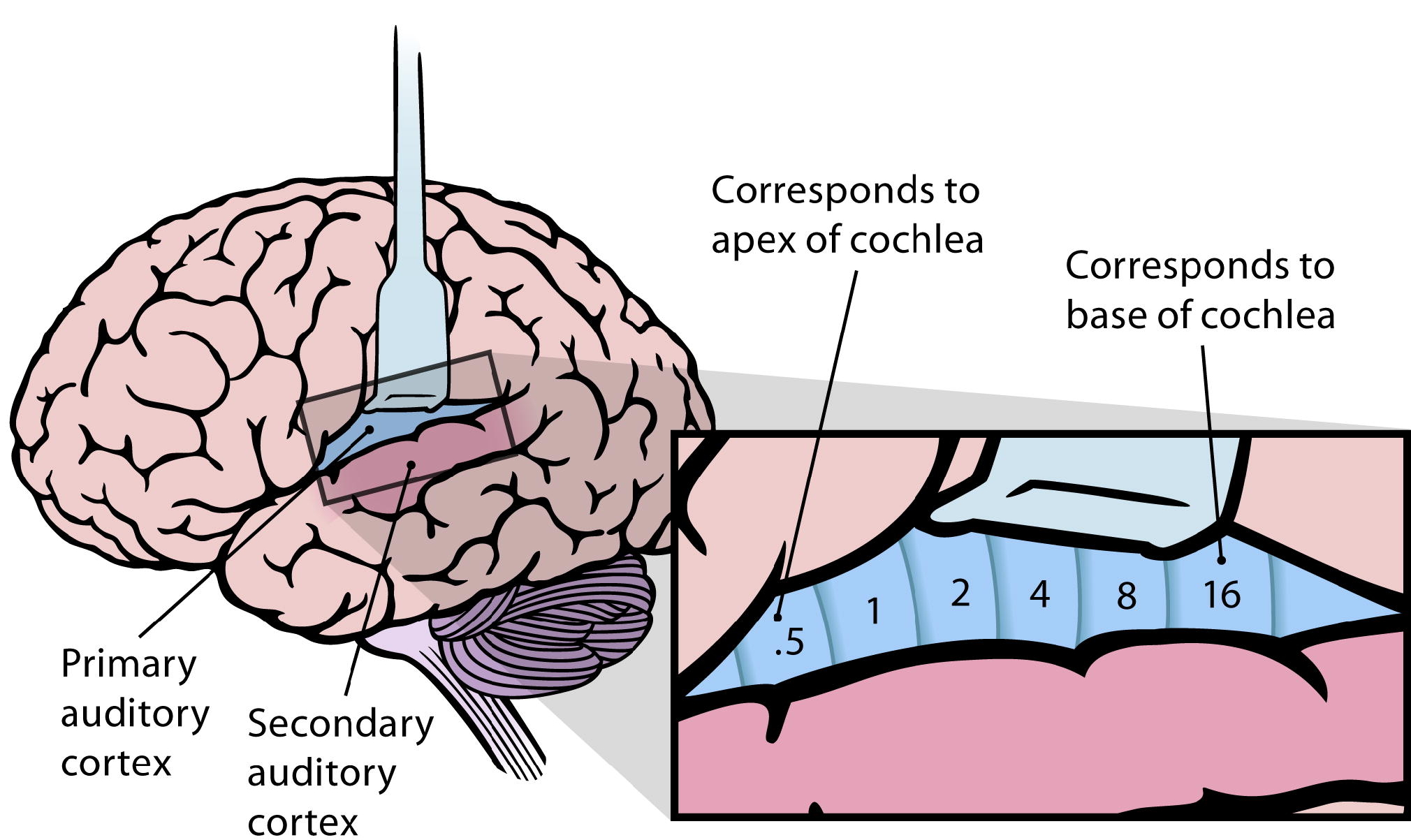
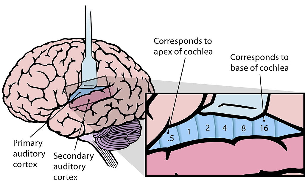
English: (B) Lateral view of the human brain, with the auditory cortex exposed. The primary auditory cortex contains a topographic map of the cochlear frequency spectrum (shown in kilohertz). (Redrawn from Figure 12.15A in [11].) |
| Date |
|
| Source | |
| Author |
|
| This is a retouched picture, which means that it has been digitally altered from its original version. Modifications: Isolated subfigure B. The original can be viewed here: 10.1371 journal.pbio.0030137.g001-L.jpg:
|
I, the copyright holder of this work, hereby publish it under the following licenses:
This file is licensed under the Creative Commons Attribution 2.5 Generic license.
- You are free:
- to share – to copy, distribute and transmit the work
- to remix – to adapt the work
- Under the following conditions:
- attribution – You must give appropriate credit, provide a link to the license, and indicate if changes were made. You may do so in any reasonable manner, but not in any way that suggests the licensor endorses you or your use.
You may select the license of your choice.
Original upload log
This image is a derivative work of the following images:
- File:10.1371_journal.pbio.0030137.g001-L.jpg licensed with Cc-by-2.5, Cc-by-2.5
- 2009-02-12T04:06:25Z Mike.lifeguard 2020x2480 (483539 Bytes) {{Information |Description={{en|1=(A) The human ear and frequency mapping in the cochlea. The three ossicles incus, malleus, and stapes transmit airborne vibration from the tympanic membrane to the oval window at the base of
Uploaded with derivativeFX
File history
Click on a date/time to view the file as it appeared at that time.
| Date/Time | Thumbnail | Dimensions | User | Comment | |
|---|---|---|---|---|---|
| current | 04:12, 12 February 2009 | | 2,020 × 1,203 (667 KB) | Mike.lifeguard | {{Information |Description={{en|1=(B) Lateral view of the human brain, with the auditory cortex exposed. The primary auditory cortex contains a topographic map of the cochlear frequency spectrum (shown in kilohertz). (Redrawn from Figure 12.15A in [11].) |
File usage
The following page uses this file:
Global file usage
The following other wikis use this file:
- Usage on ru.wikiversity.org